
|
Fig. 1
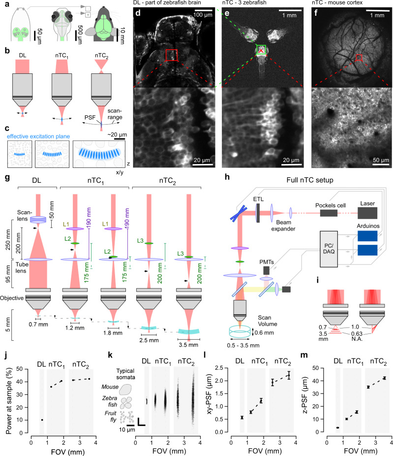
a Drosophila (left), larval zebrafish (centre) and adult mouse schematics (right) with central nervous system highlighted (green) to illustrate size differences. Insets next to the mouse for size-comparison on the same scale. b Optical configurations of standard diffraction limited (DL, left) 2P setup with parallel laser beam entering objective’s back aperture. Right, non-telecentric (nTC, middle, right) configurations use a diverging laser beam instead. As a result, the field of view and focal distance are expanded, and the point spread function (PSF) elongates. These effects scale with the angle of divergence (compare nTC1 and nTC2). c Typical neuronal somata in species shown in (a), as interrogated by 2P setups shown in (b), respectively. d In vivo 7 dpf larval zebrafish (HuC:GCaMP6f) imaged with a Sutter-MOM DL setup at full field of view (top) and when zoomed in to reveal individual neuronal somata (bottom) as indicated. e same zebrafish as (d), next to two further zebrafish imaged using nTC2 configuration at maximal field of view (top). Zooming in to the same area as in (d, bottom) reveals cellular detail (e, bottom). f In vivo adult mouse cranial window over somatosensory cortex imaged with nTC2 maximal field of view (top) and when zoomed in as indicated (bottom). g Left: Optical configuration of a standard DL setup with collimation system consisting of a scan lens and a tube lens to set-up an infinity collimated laser beam at the objective’s back aperture. Effective refractive power and relative distances of lenses indicated. The intermediary focal point (IFP) is behind the scan lens (arrowhead). Middle: nTC1 configuration replaces the scan lens with two plano-convex lenses (L1,2). The relative position of L2 to the tube lens defines the position of the IFP, which is now further along the laser path. As a result, the field of view can be expanded to between 1.2 and 1.8 mm. Right: nTC2 configuration using a single plano-convex lens (L3) allows FOV expansion to 2.5–3.5 mm. h complete nTC setup, including an ETL positioned in front of the scan mirrors for rapid axial-scanning. PMTs, Photomultipliers. i FOV expansion under nTC combines two effects: Increased focal distance (left) and reduced numerical aperture (N.A., right), which together yield a larger effective focal plane and enlarged PSF. j Power at sample measured for all configurations, expressed as a percentage of the power that reaches the scanning mirrors. k point spread functions (PSFs) measured for all configurations, with size of typical neuronal somata of different species indicated. All scale-bars 10 µm. l, m, lateral (l) and axial (m) spread of the PSFs quantified. Errors in mean ± s.d., n = 20 experimentally independent samples per measurement. Data leading to j, l, m in Source data file.