
|
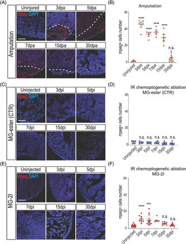
Fig. 5 Macrophages are activated after cardiomyocyte chemoptogenetic ablation. A, Mpeg and DAPI staining before and at multiple time points after ventricular apex resection. White dashed lines indicate injury area. B, Quantification of mpeg+ cells inside the injury after amputation. Between 3- and 15 dpa, macrophages were detected inside the injured area. By 30 dpa, the number of macrophages were similar to the uninjured hearts. At least n = 3 for each group. ****P < .0001 One-way ANOVA. C, Mpeg and DAPI staining in Tg(myl7:fapdl5-cerulean) hearts before and after injection of control MG-ester and near IR light exposure. D, Quantification of Mpeg positive cells in the ventricle. No differences were noted at different time points after MG-ester injection. E, Mpeg and DAPI staining before and after cardiac chemoptogenetic ablation. F, Graph with the number of Mpeg+ cells per ventricular area. Chemoptogenetic ablation causes accumulation of macrophages inside the ventricle between 3- and 7 dpi. At 15- and 30 dpi, the number of macrophages is comparable to control. At least n = 4 for each group. *P < .05; ***P < .001; ****P < .0001; one-way ANOVA. Scale bar = 100 μm