
|
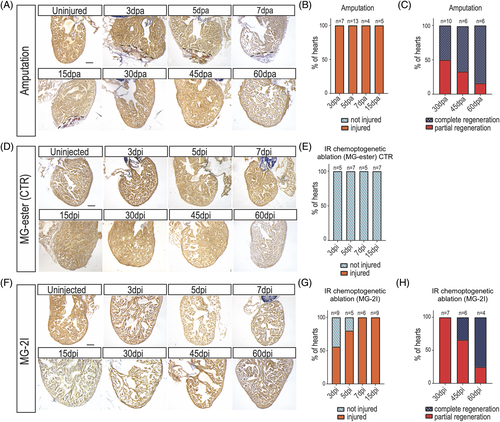
Fig. 1 Zebrafish regenerate after cardiomyocyte chemoptogenetic ablation. A, AFOG staining in WT adult zebrafish hearts before and after ventricular apex amputation. (dpa) = days post amputation. Black dashed lines delineate injured area. B, Graph representing the percentage of hearts that were injured (orange) at multiple time points. Percentages were calculated upon sectioning and AFOG staining. 100% of the hearts undergoing amputation were injured, showing the efficiency of the surgery. C, Graph reporting the percentage of hearts that were completely (blue) or partially (red) regenerated at 30-, 45-, and 60 dpa. At 30 dpa, 50% of the fish were completely regenerated, while at 60 dpa almost all the fish showed complete regeneration. D, AFOG staining of Tg(myl7:fapdl5-cerulean) hearts after injection of control MG-ester and IR light treatment at several time points. (dpi) = days post injection. Hearts appeared normal at every time point studied. Damage was determined as fraction of myocyte area of the total ventricular area. E, Graph showing that upon injection of control MG-ester and IR light treatment. F, AFOG staining of Tg(myl7:fapdl5-cerulean) hearts after injection of MG-2I and IR light treatment at several time points. Visible loss of cardiomyocytes was detected as early as 3 dpi. G, Graph representing the percentage of hearts that were injured (orange) or not injured (light blue) at several time points after MG-2I injection. At 3 dpi only 50% of the hearts showed injury, but by 7 dpi, all the hearts showed injury. H, Graph representing the percentage of hearts that show complete or partial regeneration at later time points. Total number of hearts analyzed are indicated. Scale bars = 100 μm