IMAGE

Figure 4

ID
ZDB-IMAGE-211129-62
Source
Figures for Su et al., 2021
Image
Figure Caption

Figure 4

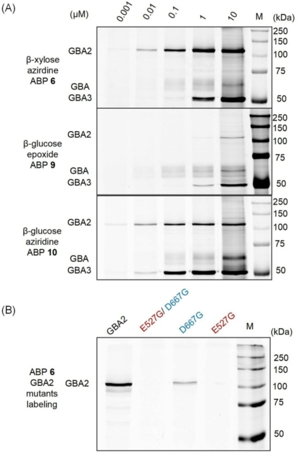
(A) ABP labeling of β‐d‐glucosidases. A lysate of HEK293T cells expressing human GBA, GBA2 and GBA3 was incubated with indicated ABPs (6, 9 or 10) for 30 min at pH 6.0. Fluorescently labeled proteins were visualized after SDS‐PAGE. (B) Labeling with ABP 6 of wild type or mutant GBA2 (E527G nucleophile mutation and D667G acid/base mutation) expressed in HEK293T cells.

Acknowledgments
This image is the copyrighted work of the attributed author or publisher, and ZFIN has permission only to display this image to its users. Additional permissions should be obtained from the applicable author or publisher of the image. Full text @ Chembiochem