
|
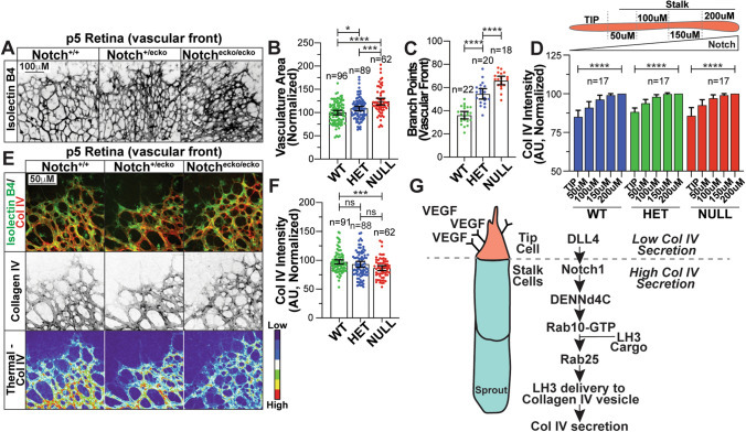
Fig. 7 Notch1 deficient mouse retinas have reduced collagen IV. (a) Representative images of wild-type (WT; Cdh5-PAC-CreER+/−; Notch+/+), Notch EC knockout(ecko)/− (HET, Cdh5-PAC-CreER+/−; Notchflox/+), or Notchecko/ecko (NULL, Cdh5-PAC-CreER+/−; Notchflox/flox) retinas harvested at P5 and stained for isolectin B4 (IB4). (b) Graph of vasculature area for vessels between indicate genotypes. (c) Graph of number of branch points at vascular front. (d) Top- schematic of Col IV measurements taken on sprouts. Graph of collagen IV (Col IV) intensity starting at the vascular front and measured back every 50 μm between indicated groups. Intensities are normalized to Col IV levels at 200 μm for each group. (e) Representative images of retinas harvested at P5 and stained for Col IV (red), DNA (blue), and IB4 (green) to identify blood vessels between indicated genotypes. (f) Graph of Col IV fluorescence intensity across indicated groups. (g) Model- VEGF-binding at the tip cells decreases Notch activation. In stalk cells, Notch activation promotes expression of DENNd4c which activates Rab10. Active Rab10 works with Rab25 to traffic Lysyl hydroxylase 3 (LH3) to Col IV-containing vesicles, allowing for secretion of Col IV into the extracellular space. For all experiments, data represented as mean ± 95% confidence intervals. N number of measurements. In all conditions, 6 or more mice were used per group. Black bars indicate comparison groups with indicated p-values. All p-values are from two-tailed Student’s t-test from duplicate experiments. *p ≤ 0.05; **p ≤ 0.01; ***p ≤ 0.001; ****p ≤ 0.0001; ns, not significant