
|
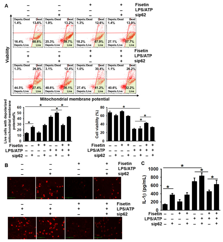
Figure 6
Transient knockdown of p62 aggravates the fisetin-induced stabilization of mitochondrial membrane potential. BV2 microglial cells were transiently transfected with p62 siRNA (sip62) for 48 h and subsequently treated with 1 µg/mL LPS for 2 h, followed by stimulation with 1 mM ATP (LPS/ATP) for 2 h. (A) The population of cells with depolarized mitochondrial membrane potential was measured using a Muse MitoPotential Kit. The total population of live cells with depolarized mitochondrial membrane potential (bottom left) and cell viability (bottom right) is depicted. (B) In a parallel experiment, cells were stained using 2 µM MitoSOX Red. Images of cells were captured using a CELENA S Digital Imaging System. (C) Extracellular level of IL-1β were quantified using ELISA after 48 h of stimulation of LPS/ATP. The results indicate the mean ± standard error median (SEM), and is representative of the results obtained from three independent experiments.* p < 0.05.