IMAGE

Fig. 1.

ID
ZDB-IMAGE-210821-31
Source
Figures for Hosseini et al., 2021
Image
Figure Caption

Fig. 1.

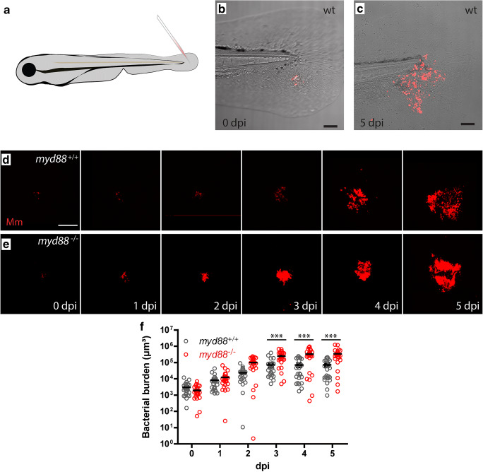
Granuloma development and morphology in the tail fin of zebrafish larvae. aMm injection in the tail fin generates single granuloma. b,c Infected tail fin of the same larva with fluorescently labelled Mm (red) at 4 h post infection (b) and at 4 days post infection (c) showing the localized development of the early granuloma structures. d,e Representative images of larvae showing the increase of Mm infection (red) and development of granuloma structures in the wild type (d) and myd88-/- larvae (e). f Bacterial burden in the myd88+/+ (black) and myd88-/- (red) larvae. The data (mean ± SEM) were analysed using analysis of variance (ANOVA); Bonferroni’s multi comparison post-test was performed on myd88 wild type and mutant larvae at each time point (***p < 0.0001, n > 20 larvae per time point). The scale bars represent 100 μm

Acknowledgments
This image is the copyrighted work of the attributed author or publisher, and ZFIN has permission only to display this image to its users. Additional permissions should be obtained from the applicable author or publisher of the image. Full text @ Virchows Arch