IMAGE

Fig 5

ID
ZDB-IMAGE-210821-12
Source
Figures for Farías-Serratos et al., 2021
Image
Figure Caption

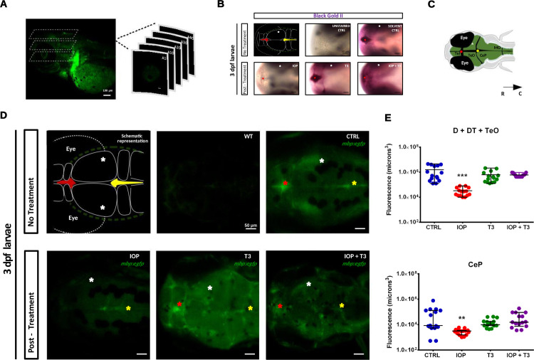
Fig 5 IOP and T3 rescue treatment in <italic toggle='yes'>mbp</italic>:<italic toggle='yes'>egfp</italic> transgenic zebrafish larvae at 3 dpf.

(A) Lateral scan of the head using confocal microscopy and generating multiple 3D optical sections; scale bar: 100 μm. (B) Representative photomicrograph of zebrafish larvae with dorsal view and R-C orientation, whole-mount Black-Gold II staining in D, DT and at 3 dpf (wild-type, CTRL, IOP, T3 and IOP + T3). scale bar: 50 μm. (C) Schematic drawing with dorsal view of the head; the asterisks represent signal in the habenula and between the telencephalon (Tel) and tectum opticum (TeO) (red); signal within the TeO (white) and signal in the dorsal Tectum and between TeO and cerebellar plate (CeP) (yellow). The black arrow represents the orientation of the larvae, rostral-caudal (R-C). (D) The images are the representation of the maximum intensity projection of the entire set of Z-stacks: uninjected CTRL (WT), CTRL, larvae treated with IOP 0.5 μM, T3 10 nM or IOP 0.5 μM + T3 10 nM; scale bar: 50 μm. (E) Graphs depicting the volume of the myelinated area in voxels, Data are shown as individual values and mean ± SEM (approximately n = 15 larvae/group). Statistical analysis was performed with one-way ANOVA coupled with Tukey’s multiple comparison test with respect to the control group. **p< 0.01 and ***p< 0.001.

Acknowledgments
This image is the copyrighted work of the attributed author or publisher, and ZFIN has permission only to display this image to its users. Additional permissions should be obtained from the applicable author or publisher of the image. Full text @ PLoS One