IMAGE

Fig. 6

ID
ZDB-IMAGE-210519-21
Source
Figures for Zhang et al., 2021
Image
Figure Caption

Fig. 6 Tumoral hypoxia imaging in mice.

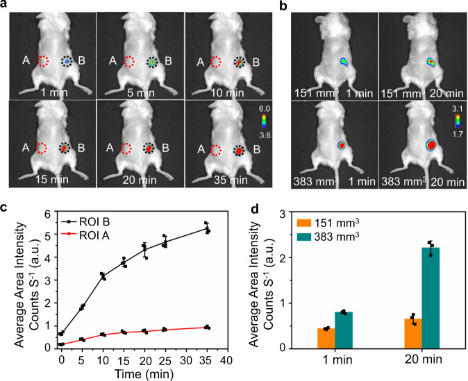
Optical imaging of human breast MCF-7 tumor-xenografted mice injected with HDSF (20 μM, 50 μL). a Images (overlaps of fluorescence and bright-field images) of a mouse underwent subcutaneously (ROI A) and intratumoral (ROI B) HDSF-injection recorded at different time post injection; b images of mice bearing tumors of different size (~151 and 383 mm3) recorded at first or 20th min post intratumoral HDSF-injection; c temporal profile of fluorescence in ROIs A and B in (a); d histogram of fluorescence in tumors in (b). Data were presented as mean ± SD. The results are representative of three independent experiments. λex, 660 nm; λem, 710 nm. Source data are available as a Source data file.

Acknowledgments
This image is the copyrighted work of the attributed author or publisher, and ZFIN has permission only to display this image to its users. Additional permissions should be obtained from the applicable author or publisher of the image. Full text @ Nat. Commun.