IMAGE

Fig 6

ID
ZDB-IMAGE-210422-19
Source
Figures for Sutton et al., 2021
Image
Figure Caption

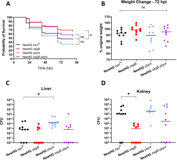
Fig 6 Analysis of growth and virulence of NewHG <italic>sagB pbp4</italic>.

(A) Approximately 1500 CFU of bacteria (mutant or wildtype) was injected into the circulation valley of LWT zebrafish embryos around 30 hpf. A survival curve was produced comparing the virulence of parental NewHG (SJF 3663, black line) to: NewHG sagB::kan (SJF 4912, red line), NewHG pbp4::ery (SJF 5103, blue line) and NewHG sagB::kan pbp4::ery (SJF 5147, purple line). (3 repeats, n>20, * p = 0.0157). Mice (n = 10) were injected intravenously with approximately 1x10⁷ CFU S. aureus NewHG kanR (WT, SJF 3680), NewHG sagB::kan (SJF 4912), NewHG pbp4::ery (SJF 5103) or NewHG sagB::kan pbp4::ery (SJF 5147). (B) Weight loss 72 hpi and CFUs recovered from (C) livers (* p = 0.0119) and (D) kidneys (* p = 0.0055). Groups were compared using a Mann-Whitney U test (NewHG kanR–black circles, NewHG sagB::kan–red squares, NewHG pbp4::ery blue diamonds, NewHG sagB::kan pbp4::ery–purple triangles).

Figure Data
Acknowledgments
This image is the copyrighted work of the attributed author or publisher, and ZFIN has permission only to display this image to its users. Additional permissions should be obtained from the applicable author or publisher of the image. Full text @ PLoS Pathog.