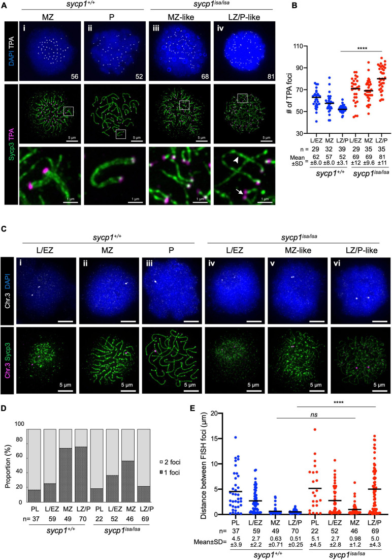
IMAGE

FIGURE 4

ID
ZDB-IMAGE-210414-24
Source
Figures for Imai et al., 2021
Image
Figure Caption

FIGURE 4

Transient pairing of homologs at chromosome ends in sycp1 mutant spermatocytes. (A) costaining of telomeres (TPA) and Sycp3 in sycp1+/+ (i and ii) and sycp1isa/isa (iii and iv) spermatocytes. The arrowhead indicates complete axis formation between two telomeres, and the arrow indicates paired telomere foci in LZ/P-like sycp1isa/isa spermatocytes. Numbers in the top panels indicate counts of TPA foci. Regions indicated in white rectangles are shown at a higher magnification at the bottom. (B) quantification of telomere polyamide (TPA) foci in sycp1+/+ and sycp1isa/isa spermatocytes. Black bars indicate means. Statistical significance was examined by a two-tailed Mann-Whitney test (****P < 0.0001; exact P value). (C) fish staining of a peritelomeric locus on chromosome 3 in sycp1+/+ (i to iii) and sycp1isa/isa (iv to vi) spermatocytes. (D) Proportion of nuclei with one or two FISH foci in sycp1+/+ and sycp1isa/isa spermatocytes. (E) quantification of distances between FISH foci of a peritelomeric locus on chromosome 3 in sycp1+/+ and sycp1isa/isa spermatocytes. Distances between FISH foci observed in Figure 4D. For nuclei with only one FISH focus, the radius of the FISH-stained area was considered the distance between two FISH foci. Black bars indicate means. Statistical significance was examined by a two-tailed Mann-Whitney test (****P < 0.0001, ns, not significant; exact P value).

Acknowledgments
This image is the copyrighted work of the attributed author or publisher, and ZFIN has permission only to display this image to its users. Additional permissions should be obtained from the applicable author or publisher of the image. Full text @ Front Cell Dev Biol