Image
Figure Caption

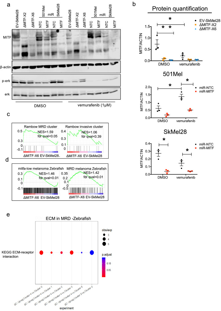
Figure 6—figure supplement 2 MITF-KO cells mimic MRD in melanoma.

(a, b) Western blot analysis and quantification of MITF, ERK and p-ERK in EV-SkMel28, ΔMITF-X2, ΔMITF-X6 and miR-NTC, miR-MITF in SkMEL28 and 501Mel cell lines treated with DMSO or vemurafenib (1 µM) for 24 hr. Actin was used as loading control. (c, d) GSEA analysis using DEGs of ∆MITF-X6 vs. EV-SkMel28 cells on Rambow MRD and invasive gene signatures and MRD signature from Zebrafish and mitfalow melanoma tumors. (e) Gene enrichment analysis plotted using Cluster profiler of single cell clusters obtained from melanoma tumors in zebrafish.

Acknowledgments
This image is the copyrighted work of the attributed author or publisher, and ZFIN has permission only to display this image to its users. Additional permissions should be obtained from the applicable author or publisher of the image. Full text @ Elife