
|
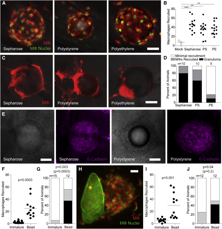
Figure 4
Chemically Inert Beads Induce Epithelioid Granulomas
(A) Representative confocal images of macrophages recruited 6 hpi of sepharose, polystyrene, or polyethylene microspheres into the HBV of transgenic zebrafish larvae carrying red-fluorescent macrophages with green nuclei.
(B) Enumeration of macrophages recruited to these microspheres in multiple animals.
(C) Representative confocal images of granulomas formed around the three types of microspheres 5 dpi into the HBV of transgenic larvae carrying the transgene for red-fluorescent macrophages (without green nuclei).
(D) Stages of macrophage recruitment to microspheres 5 dpi into HBV of multiple larvae.
(E) Brightfield (panels 1 and 3) and fluorescence confocal (panels 2 and 4) microscopy of sepharose and polystyrene bead granulomas following immunofluorescence staining with the E-cadherin antibody.
(F and G) Macrophage recruitment to immature eggs or microspheres implanted into the HBV at 6 hpi (F) and 5 dpi (G).
(H–J) Macrophage recruitment following co-implantation of an immature egg and a polystyrene microsphere into the HBV of larvae transgenic for red-fluorescent macrophages with green nuclei. (H) Representative confocal image of an immature egg next to a microsphere.
(I and J) Quantification of macrophage recruitment at 6 hpi (I) and at 5 dpi (J). Scale bars, 25 μm.
(B, F, and I) Horizontal bars, means. (D, G, and J) n, number of animals. Statistics, one-way ANOVA (B), unpaired (F), and paired (I) Student’s t tests and Fisher’s exact test comparing granulomas (black bars) or granuloma formation with macrophage recruitment (black and gray bars, in parentheses) (G–J) Experiments in (E) and (F–J) were performed once. (A–D) are representative of three experiments. Also see