IMAGE

FIGURE 7

ID
ZDB-IMAGE-210103-29
Source
Figures for Wu et al., 2020
Image
Figure Caption

FIGURE 7

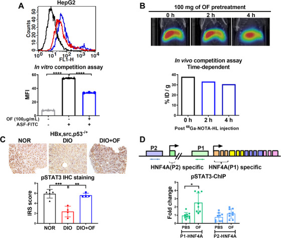
Oligo‐fucoidan binds to ASGR1/2 in hepatoma cells, enhances pSTAT3 in hepatic tissues of [HBx,src,p53−/+] transgenic fish, and enriches pSTAT3 binding to P1‐HNF4A promoter in hepatoma cells. A, Flow cytometry profiles of HepG2 cells. The mean fluorescence intensity (MFI) profiles of HepG2 cells with OF treatment. The asialofetuin‐FITC (ASF‐FITC) signals were presented in the control (black), without OF co‐treatment (blue) and with OF co‐treatment. B, The reduction of 68Ga‐NOTA‐HL radioactivity intensity in a time‐dependent manner was depended on the administration of OF (N = 1). Relative radioactive intensity of 68Ga‐NOTA‐HL in vivo between different time courses. Black bar indicates post‐68Ga‐NOTA‐HL injection 0 hour, blue and red bar denote post‐68Ga‐NOTA‐HL injection 2 hours and 4 hours, respectively. C, Immunohistochemistry staining of pSTAT3 in [HBx,src,p53−/+] transgenic fish fed with normal diet (NOR), diet‐induced obesity (DIO) or treated with oligo‐fucoidan (DIO+OF). Semi‐quantitative analysis of pSTAT3 staining in hepatic tissues from [HBx,src,p53−/+] transgenic zebrafish. Statistical significance was calculated by t‐test: **P ≤ .01, ***P ≤ .001. D, The expression fold of chromatin immunoprecipitation (ChIP) of pSTAT3 binding to P1‐HNF4A promoter in HepG2 cells was enriched by OF treatment. Statistical significance was calculated by t‐test: *P ≤ .05, **P < .01, ***P ≤ .001; **** P ≤ .0001.

Figure Data
Acknowledgments
This image is the copyrighted work of the attributed author or publisher, and ZFIN has permission only to display this image to its users. Additional permissions should be obtained from the applicable author or publisher of the image. Full text @ Clin Transl Med