IMAGE

Figure 12

ID
ZDB-IMAGE-201229-91
Source
Figures for Candiani et al., 2020
Image
Figure Caption

Figure 12

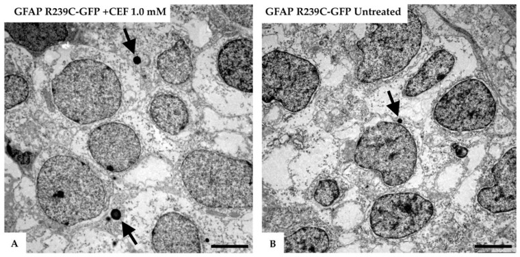
Electron micrographs of the telencephalon of 3 dpf zebrafish embryos expressing GFAP R239C-GFP.(A) The formation of lysosomes (arrows) was confirmed in the electron micrographs of the telencephalic region from a 3 dpf old zebrafish, injected with GFAP R239C-GFP plasmid and treated with 1.0 mM ceftriaxone for 48 h. (B) In contrast, in mutant untreated embryos, fewer and smaller lysosomes were seen at this embryonic stage. Images were captured with Zeiss EM10 or a LEO 912AB transmission electron microscope. Scale bar: 2.5 µm.

Acknowledgments
This image is the copyrighted work of the attributed author or publisher, and ZFIN has permission only to display this image to its users. Additional permissions should be obtained from the applicable author or publisher of the image. Full text @ Genes (Basel)