
|
Figure 1
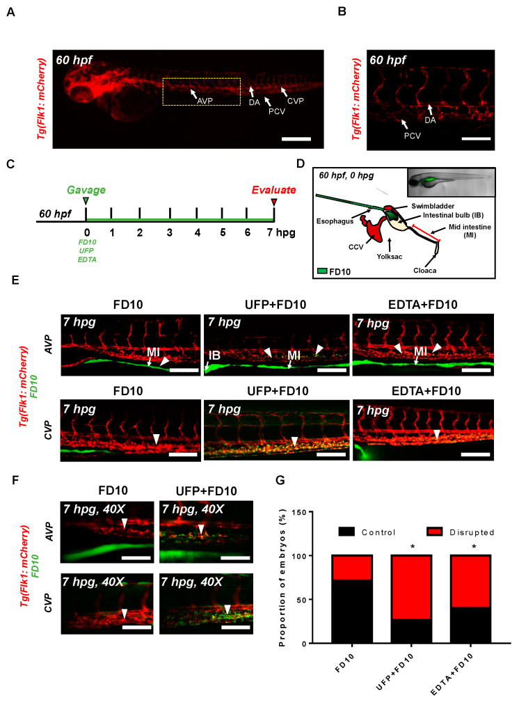
Acute ultrafine particulate matter (UFP) ingestion disrupts intestinal epithelial barrier integrity. Transgenic Tg(flk1: mCherry) zebrafish embryos at 60 hpf were micro-gavaged with FITC-conjugated dextran (FD10, 10 kDa). (A,B) Anatomy of endothelial vasculature in the Tg(flk1: mCherry) embryo. DA: dorsal aorta; PCV: posterior caudal vein; AVP: anterior venous capillary plexus; CVP: caudal vein capillary plexus; Scale bar: 32 μm. (C) Experimental design: At 2 dpf, embryos were randomly chosen for micro-gavage with FD10 solution with or without UFP or EDTA at 20 mM. Intestinal barrier integrity and translocation of FD10 to vascular endolumen (flk1+) were evaluated at 7 h post gavage (hpg). (D) A schematic representation of micro-gavage in an embryonic gastrointestinal (GI) tract. FD10 solution was micro-gavaged in the intestinal bulb without disrupting the esophagus, swim bladder and yolk sac. (E) Representative images of the AVP and CVP at 7 hpg. In FD10 gavaged-controls, FD10 remained only in the intestinal bulb and mid-intestine. In contrast, co-gavaging FD10 with UFP or ethylenediaminetetraacetic acid (EDTA) accumulated FD10 in the AVP and CVP (white arrow heads). Scale bar: 20 μm. (F) Magnified view of the AVP and CVP. Scale bar: 20 μm. (G) Percentage of embryos exhibiting endoluminal FD10 fluorescence (* p < 0.05 vs. FD10, n = 10 per group).