IMAGE

Figure 1

ID
ZDB-IMAGE-200805-7
Genes
Antibodies
Source
Figures for Grivas et al., 2020
Image
Figure Caption

Figure 1

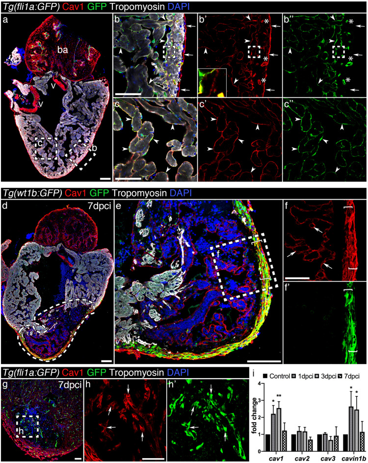
Caveolin-1 is expressed in the endothelium, endocardium and epicardium of the intact and injured adult zebrafish heart. (a) Immunofluorescence staining of Cav1 and Tropomyosin (cardiomyocytes) in an intact Tg(fli1a:GFP) heart. ba, bulbus arteriosus; v, valves. (bc′′) Cav1 immunoreactivity in the epicardium (arrows) overlaps with GFP in the endothelium (asterisks) and endocardium (arrowheads). Cav1 is also expressed in the zone between the cortical and trabecular cardiomyocytes (insert in b′). (df′) Cav1 immunostaining in 7 dpci Tg(wt1b:GFP) heart. The dashed area in (d) marks the injured area; Cav1 is expressed in the activated epicardium (ef′ brackets) and endocardium (f, arrows) upon injury. (gh′) Immunolabelling of Cav1 in a 7 dpci Tg(fli1a:GFP) heart. (hh′) Arrows indicate Cav1+ endocardial cells. Scale bars: 100 μm in (a), (d), (e) and 50 μm in other panels. (i) qPCR analysis of caveolae-related genes during regeneration. Mean ± s.d., Brown-Forsythe and Welch ANOVA tests, *P < 0.05, **P < 0.01.

Figure Data
Acknowledgments
This image is the copyrighted work of the attributed author or publisher, and ZFIN has permission only to display this image to its users. Additional permissions should be obtained from the applicable author or publisher of the image. Full text @ Sci. Rep.