
Image
|
Figure Caption
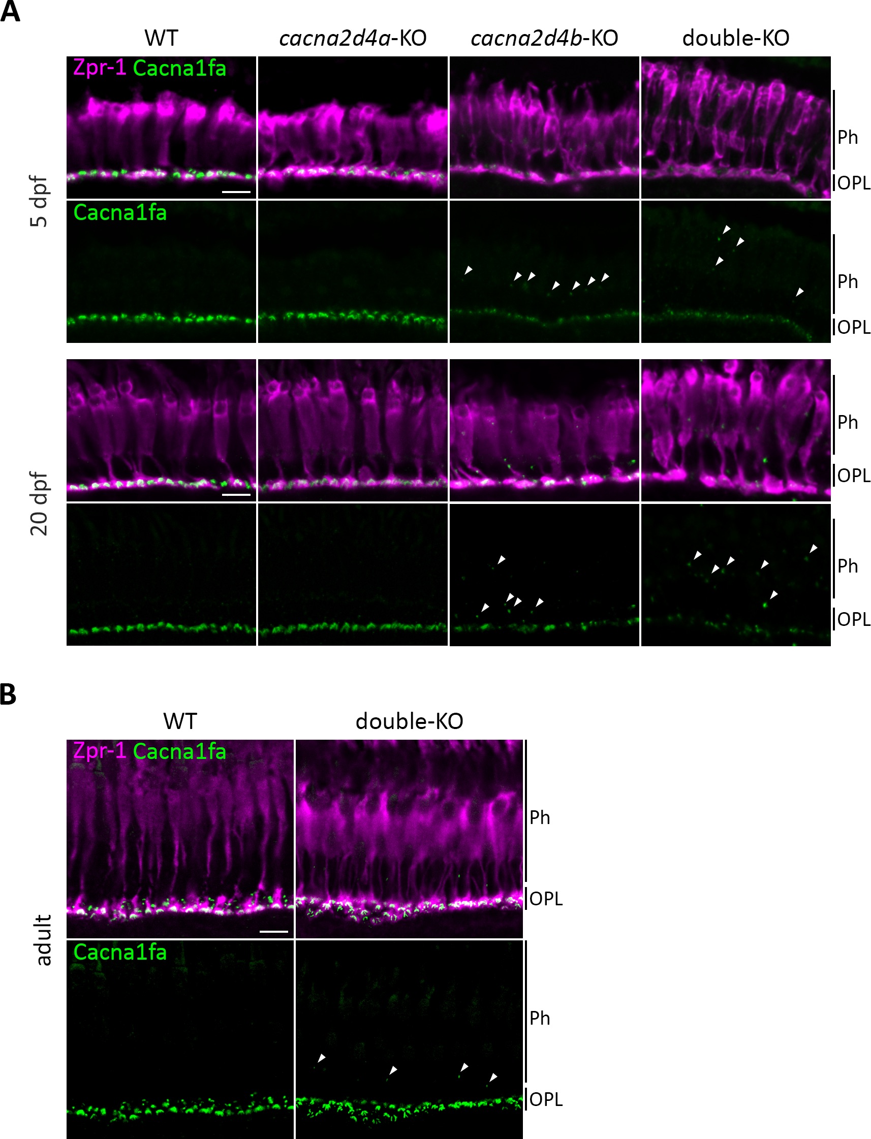
Fig. 2 Cacna1fa localization cacna2d4-KO lines. (A) Immunohistochemical analysis of Cacna1fa with Zpr-1 counterstain (red-green double cones). Cacna1fa expression is not affected in cacna2d4a-KO larvae at 5 and 20 dpf, but severely reduced in cacna2d4b-KO as well as in double-KO. In the latter two, ectopic punctate Cacna1fa staining is observed distal to the OPL (arrowheads). (B) Expression in adult double-KO is comparable to WT levels, yet ectopic puncta are visible (arrowheads). Scale bars (A, B) correspond to 10 μm and apply to all images of the respective developmental stage.
Figure Data
Acknowledgments
This image is the copyrighted work of the attributed author or publisher, and
ZFIN has permission only to display this image to its users.
Additional permissions should be obtained from the applicable author or publisher of the image.
Full text @ Invest. Ophthalmol. Vis. Sci.