IMAGE

Figure 3

ID
ZDB-IMAGE-200713-14
Source
Figures for Angileri et al., 2020
Image
Figure Caption

Figure 3

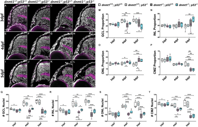
Loss of p53 function does not rescue the dnmt1−/− CMZ phenotype. AL Transverse sections of the dorsal CMZ in wildtype (A, E, I), dnmt1+/+; p53−/− (B, F, J), dnmt1−/−; p53+/+ (C, G, K), dnmt1−/−; p53−/− (D, H, L) larvae from 3 to 5dpf. Nuclei labeled with DAPI (gray) and F-actin labeled with phalloidin (magenta). MP Graphs depicting changes in retinal domain proportions over time. QT Number of nuclei in each retinal domain of dnmt1+/+; p53+/+, dnmt1+/+; p53-/, dnmt1−/−; p53+/+, and dnmt1−/−; p53−/− larvae from 3 to 5dpf. GCL ganglion cell layer, INL inner nuclear layer, ONL outer nuclear layer; CMZ ciliary marginal zone. Scale bars = 25 μm. *p < 0.05; **p < 0.005; ***p < 0.0005; ****p < 0.00005. Dorsal is up in all images.

Acknowledgments
This image is the copyrighted work of the attributed author or publisher, and ZFIN has permission only to display this image to its users. Additional permissions should be obtained from the applicable author or publisher of the image. Full text @ Sci. Rep.