IMAGE

Figure 3

ID
ZDB-IMAGE-200627-33
Source
Figures for Linnerz et al., 2020
Image
Figure Caption

Figure 3

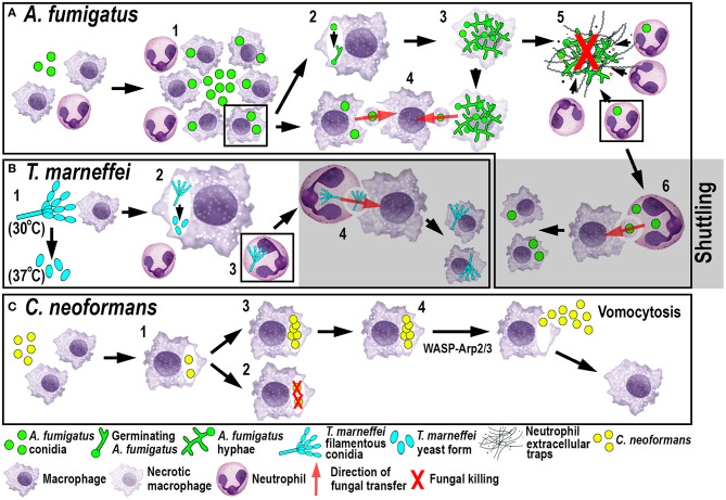
Schematic illustration of phagocyte responses to the fungal pathogens A. fumigatus(A), T. marneffei(B) and C. neoformans(C) in larval zebrafish. (A) Macrophages phagocytose A. fumigatus conidia and form tight clusters around the fungus, which inhibits fungal germination (1). Fungal germination can occur in the late phagosomes of infected macrophages (2) causing macrophage necroptosis (3). Transfer of A. fumigatus conidia can occur from infected and dying macrophages to recipient macrophages (4). Neutrophils kill A. fumigatus hyphae with their effector functions, including phagocytosis and NETosis (5). Infected neutrophils can transfer A. fumigatus to recipient macrophages through shuttling (6). (B)T. marneffei exists as filamentous conidia at 30°C and as pathogenic yeast form at 37°C (1). T. marneffei spores can transition to the yeast form within macrophages (2). Neutrophils can also phagocytose T. marneffei conidia (3) and transfer them to recipient macrophages through shuttling (4). (C) Macrophages phagocytose C. neoformans (1) where it can be killed (2) or persist and proliferate (3). WASP-Arp2/3 regulates vomocytosis and fungal dissemination (4).

Acknowledgments
This image is the copyrighted work of the attributed author or publisher, and ZFIN has permission only to display this image to its users. Additional permissions should be obtained from the applicable author or publisher of the image. Full text @ Front Immunol