
|
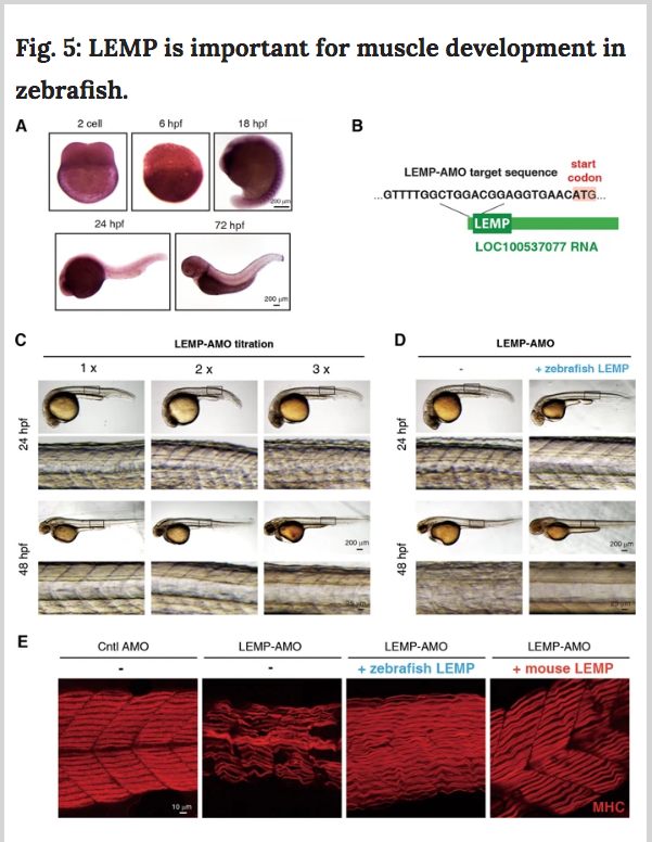
Fig. 5 a In situ hybridization to examine the localization of MyolncR4 at different developmental stages of zebrafish embryos with a specific probe targeting zLEMP. hpf, hours post fertilization. Scale bar, 200 μm. b Diagram to show the targeting sequence of the LEMP-AMO. c Representative images of zebrafish at 24 or 48-hpf after injection of different amounts of LEMP-AMO at one-cell stage. Scale bar, 200 μm or 25 μm. d Representative images of zebrafish at 24 or 48-hpf after injection of LEMP-AMO in the presence or absence of the zebrafish LEMP mRNA at one-cell stage. Scale bar, 200 or 25 μm. e MHC immunostaining to examine myofiber alignment in 48-hpf embryos injected with Cntl-AMO, LEMP-AMO, LEMP-AMO with zLEMP mRNA, or LEMP-AMO with mLEMP mRNA, respectively. Scale bar, 10 μm.