
Image
|
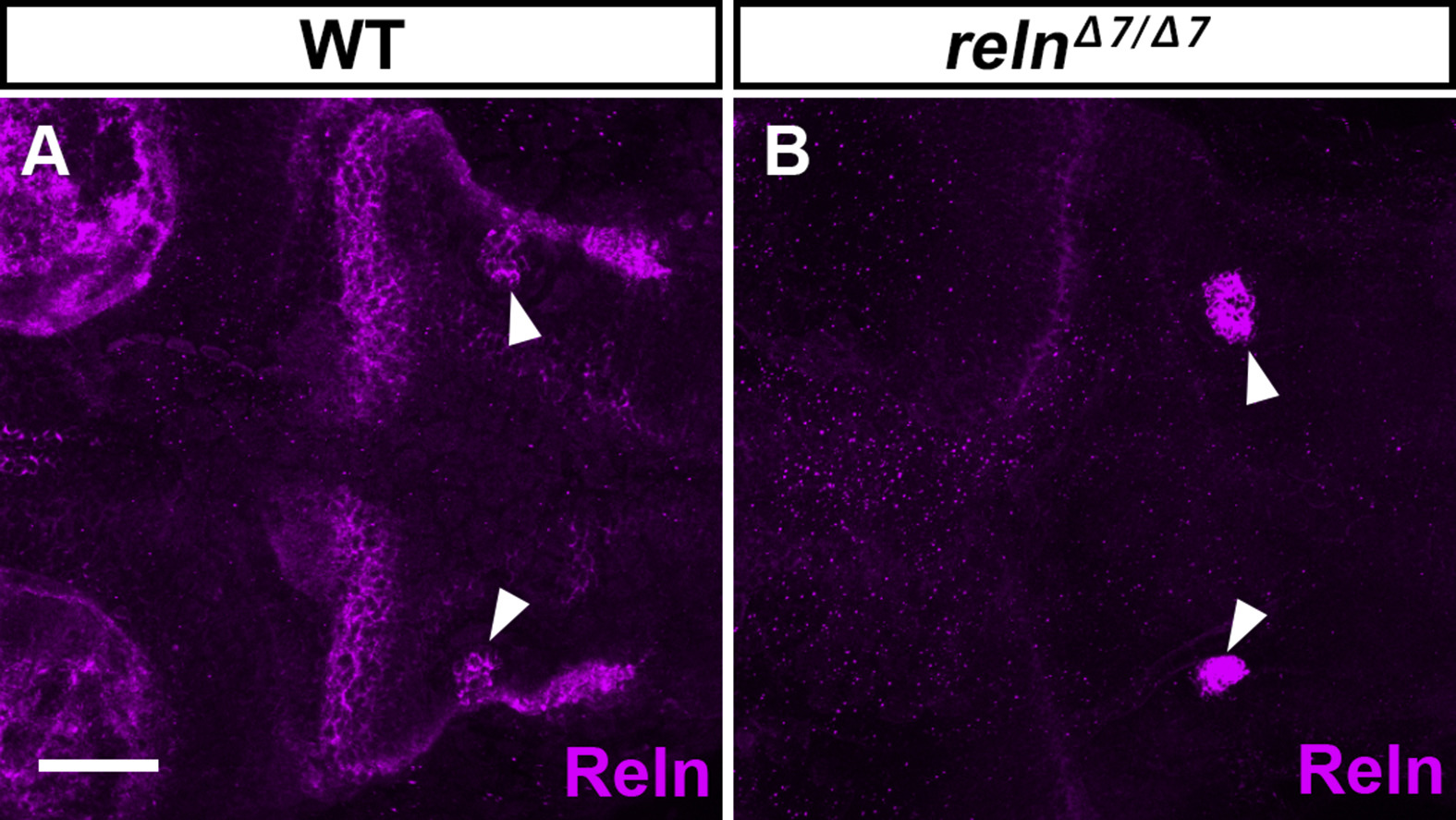
Figure Caption
Fig. S13
Specific detection of Reln protein by immunostaining. WT (A, n= 4) and relnΔ7/Δ7 (B, n= 3) larvae at 5-dpf were stained with anti-Reln (magenta) antibody. Dorsal views with anterior to the left. Typical images are shown. Note that Reln signals in and near the granule cell axons were severely decreased in the reln mutants. In some reln mutant larvae, nonspecific signals were also seen bilaterally in the hindbrain (marked by arrowheads). Scale bars: 100 μm in A.
Acknowledgments
This image is the copyrighted work of the attributed author or publisher, and
ZFIN has permission only to display this image to its users.
Additional permissions should be obtained from the applicable author or publisher of the image.
Reprinted from Developmental Biology, 455(2), Nimura, T., Itoh, T., Hagio, H., Hayashi, T., Di Donato, V., Takeuchi, M., Itoh, T., Inoguchi, F., Sato, Y., Yamamoto, N., Katsuyama, Y., Del Bene, F., Shimizu, T., Hibi, M., Role of Reelin in cell positioning in the cerebellum and the cerebellum-like structure in zebrafish, 393-408, Copyright (2019) with permission from Elsevier. Full text @ Dev. Biol.