
Image
|
Figure Caption
Fig. S5
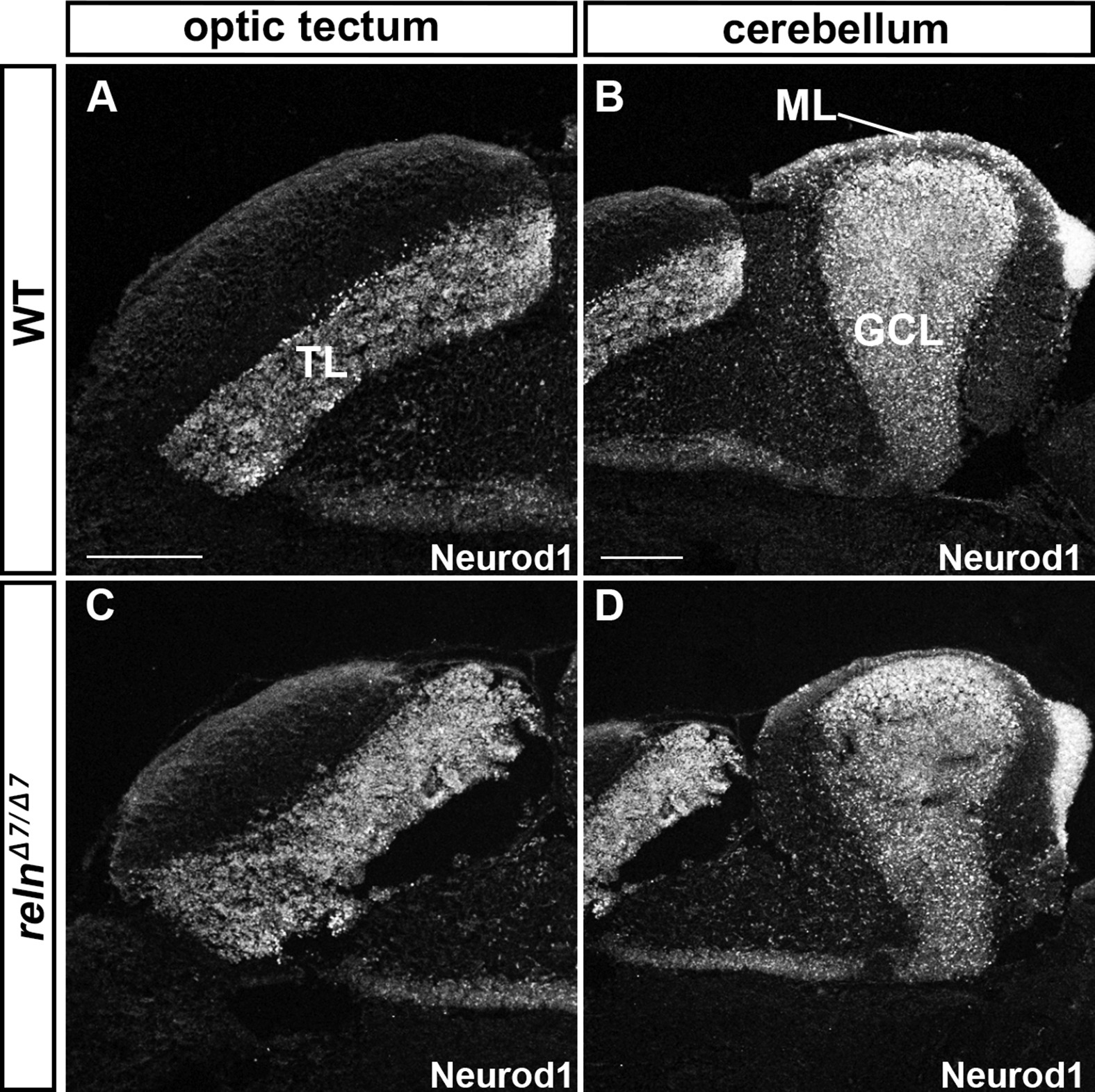
Granule cell (GC) development is not affected in reln mutants. Sagittal sections of the brain from adult (93-dpf) WT (A, B) and relnΔ7/Δ7(C, D) fish were stained with anti-Neurod1 antibodies, which mark the nucleus of immature and mature GCs in the TL, ML, and GCL. Note that the number and position of GC somata were largely unaffected in the relnmutants (n= 6 for WT, and n= 6 for relnΔ7/Δ7 mutants). The abbreviations are described in the legend of Fig. 1. Sale bars: 100 μm in A (applies to A, C); 200 μm in B (applies to B, D).
Acknowledgments
This image is the copyrighted work of the attributed author or publisher, and
ZFIN has permission only to display this image to its users.
Additional permissions should be obtained from the applicable author or publisher of the image.
Reprinted from Developmental Biology, 455(2), Nimura, T., Itoh, T., Hagio, H., Hayashi, T., Di Donato, V., Takeuchi, M., Itoh, T., Inoguchi, F., Sato, Y., Yamamoto, N., Katsuyama, Y., Del Bene, F., Shimizu, T., Hibi, M., Role of Reelin in cell positioning in the cerebellum and the cerebellum-like structure in zebrafish, 393-408, Copyright (2019) with permission from Elsevier. Full text @ Dev. Biol.