IMAGE

Fig. 5

ID
ZDB-IMAGE-200212-8
Source
Figures for Song et al., 2020
Image
Figure Caption

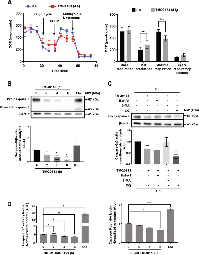
Fig. 5 Effect of TMQ0153 on cellular metabolism.

a Oxygen consumption rate (OCR) was measured by Seahorse XFp analyzer. b Caspase 8 analysis by western blot (upper panel) and the corresponding densitometric analysis (lower panel). Etoposide (Eto; 100 µM, 24 h) was used as a positive control for apoptotic caspase cleavage. c Caspase 8 analysis by western blot (upper panel) in the presence or absence of autophagy inhibitors: 40 nM baf-A1, 10 mM, 3-methyladenine (3-MA) and 75 μM chloroquine (CQ) and the corresponding densitometric analysis (lower panel). In western blot analyses, β-actin was used as a loading control. d Quantification of caspases-3/7 (left graph) and -9 activity (right graph) levels at 30 µM of TMQ0153 treatment. Etoposide (Eto; 100 µM, 24 h) was used as a positive control for apoptosis induction. All pictures are representative of three independent experiments and data represent mean (±S.D.) of three independent experiments. Statistical significance was assessed as *p < 0.05, **p < 0.01, ***p < 0.001 compared to untreated cells unless otherwise specified. One-way ANOVA (Caspase-3/7 and -9 assay); post hoc; Tukey’s test. Two-way ANOVA (mito stress test); post hoc; Sidak’s test. One-way ANOVA (western blot quantification); post hoc; Dunnett’s test.

Acknowledgments
This image is the copyrighted work of the attributed author or publisher, and ZFIN has permission only to display this image to its users. Additional permissions should be obtained from the applicable author or publisher of the image. Full text @ Cell Death Dis.