IMAGE

Figure 3

ID
ZDB-IMAGE-200131-9
Source
Figures for Caro-Astorga et al., 2020
Image
Figure Caption

Figure 3

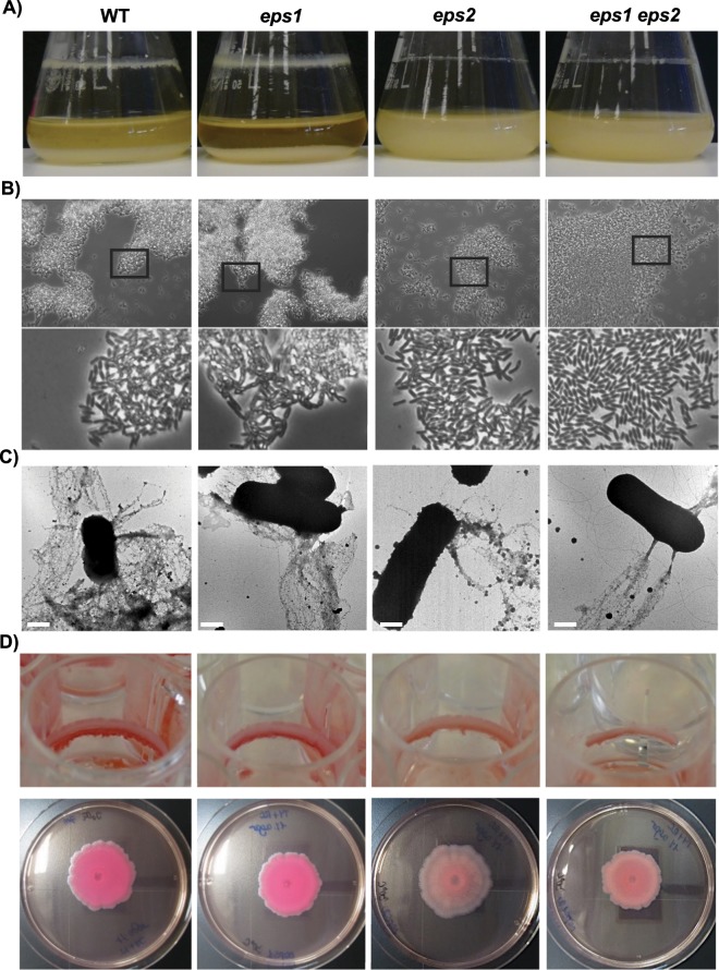
Biofilm phenotypes of the WT and eps-mutant strains. (A) Liquid cultures in TY medium incubated for 24 h at 30 °C with 150 rpm shaking showed that the WT strain yielded the largest amount of biomass attached to the walls of the flask compared to the strain mutated in the eps2 region. (B) Phase contrast micrographs of the spent medium from liquid cultures show the presence of bacterial clumps in the media from the WT and Δeps1 strains. The bottom pictures are closer views of the squared areas in the original pictures (C) Transmission electron micrographs of negatively stained cells decorated with fibers reactive to anti-TasA antibodies. (D) Congo Red biofilm assays in: Top, TY liquid medium supplemented with Congo Red and incubated without shaking at 30 °C for 6 days, and bottom, TY medium solidified with 1.5% agar supplemented with Congo Red and incubated at 30 °C for 72 h. The bars in D = 1 μm.

Acknowledgments
This image is the copyrighted work of the attributed author or publisher, and ZFIN has permission only to display this image to its users. Additional permissions should be obtained from the applicable author or publisher of the image. Full text @ Sci. Rep.