IMAGE

Figure 2

ID
ZDB-IMAGE-200129-35
Genes
Antibodies
Source
Figures for Fallata et al., 2019
Image
Figure Caption

Figure 2

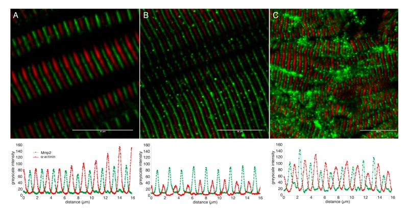
Mmp2 is localized between Z-discs in sarcomeres of embryonic and adult muscle. Confocal micrographs of skeletal muscle from 72 hpf embryos (A) or adults (B), and 500 nm thick cryosection of 72 hpf skeletal muscle (C) stained with anti-α-actinin (red) and anti-Mmp2 (green). Greyscale intensity profiles of both channels along a line drawn perpendicular to the sarcomeres are shown below each micrograph. Mmp2 immunoreactivity occurs at regularly spaced intervals precisely between the α-actinin-labeled Z-discs. Scale bars = 10 µm.

Figure Data
Acknowledgments
This image is the copyrighted work of the attributed author or publisher, and ZFIN has permission only to display this image to its users. Additional permissions should be obtained from the applicable author or publisher of the image. Full text @ Biomedicines