IMAGE

Figure 2

ID
ZDB-IMAGE-191230-991
Genes
Source
Figures for Suzuki et al., 2019
Image
Figure Caption

Figure 2

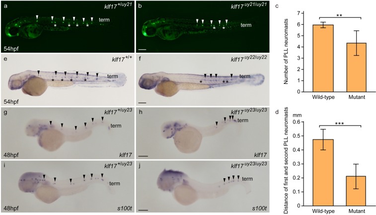
Abnormal PLL neuromast deposition in the klf17-deficient embryos. (ad) Lateral line neuromasts stained with 4-Di-2-ASP (Di-ASP). PLL neuromasts were bilaterally stained with Di-ASP. White arrowheads indicate the position of PLL neuromasts on the left side. White asterisks indicate the position of PLL neuromasts on the right side. Term indicates the position of terminal neuromasts. The number of PLL neuromasts (c) and the distance between first and second neuromasts (d) were measured in wild-type (n = 16) and the klf17-deficient embryos (n = 9). Asterisk indicates statistical significance between wild-type and the klf17-deficient embryos. **P < 0.01. ***P < 0.001. Error bars indicate standard deviation. (e,f) Alkaline phosphatase accumulation in the lateral line neuromasts. Arrowheads indicate the position of PLL neuromasts on the left side. Asterisks indicate the position of PLL neuromasts on the right side. (gj) The expression of lateral line genes, klf17 (g,h) and s100t (i,j), was examined by whole-mount in situ hybridization (WISH) at 48 hpf. (g,i) Wild-type embryos (klf17+/uy23). (h,j) klf17-deficient embryos (klf17uy23/uy23). Arrowheads indicate the position of PLL neuromasts on the left side. All pictures showed lateral view, anterior left. After taking pictures, genotyping of individual embryos was performed by genomic PCR. Scale bar, 200 μm.

Figure Data
Acknowledgments
This image is the copyrighted work of the attributed author or publisher, and ZFIN has permission only to display this image to its users. Additional permissions should be obtained from the applicable author or publisher of the image. Full text @ Sci. Rep.