IMAGE

Figure 6

ID
ZDB-IMAGE-191230-1734
Source
Figures for Simbulan-Rosenthal et al., 2019
Image
Figure Caption

Figure 6

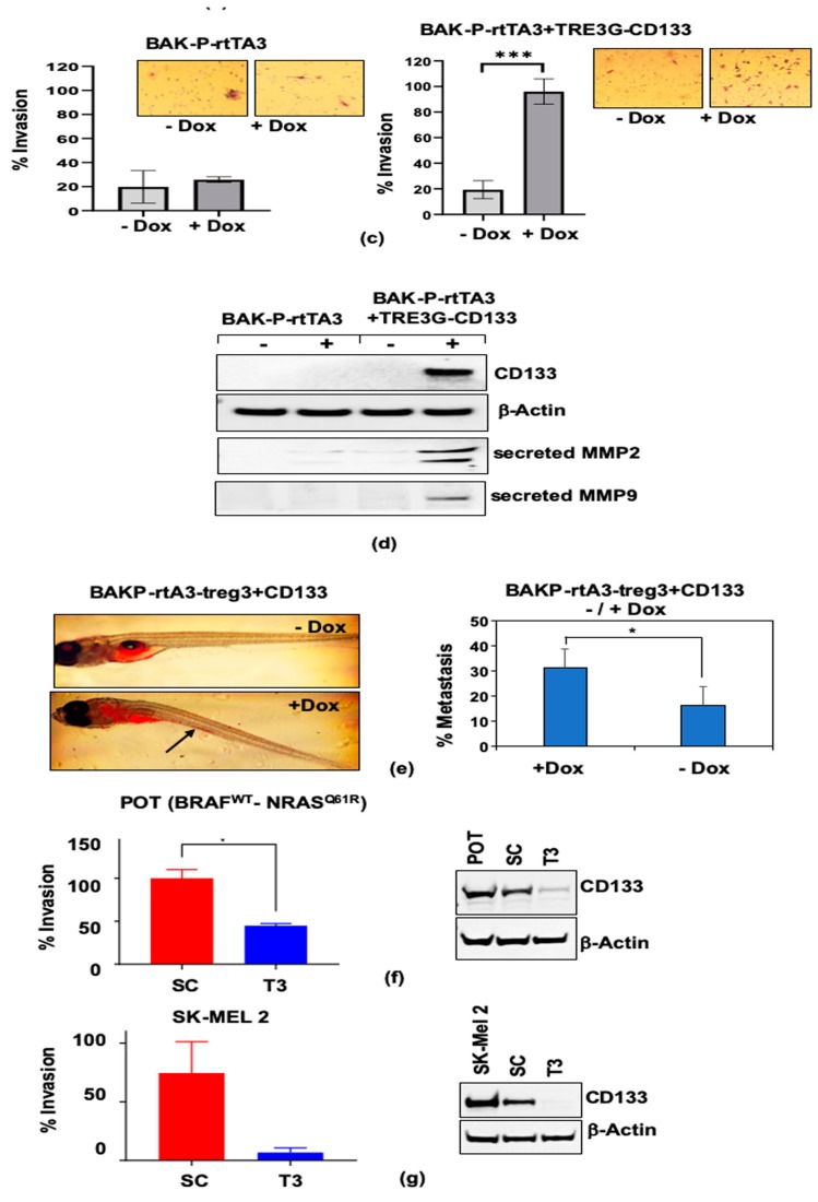
Dox-inducible CD133 expression in BAK-P cells as verified by qPCR (a) and immunoblot analysis (b). (c) Dox-inducible CD133 expression significantly increases cell invasion in transwell assays. (d) Immunoblot analysis with antibodies to CD133, MMP9, MMP2, or β-Actin for loading controls, show increased levels of CD133 as well as MP2 and MMP9 secreted by Dox-induced cells. (e) Zebrafish assays reveal enhanced metastasis in Dox-induced cells. (f,g left panels) Invasion assay results with other melanoma cell lines POT (f) and SK-MEL2 (g) CD133 CRISPR-cas9 SC versus T3 lines are consistent with results with BAK-P cells, showing that knockdown of CD133 (f,g right panels) results in decreased cell invasion. *, **, *** represent p < 0.05 and p < 0.01, and p < 0.001, respectively.

Acknowledgments
This image is the copyrighted work of the attributed author or publisher, and ZFIN has permission only to display this image to its users. Additional permissions should be obtained from the applicable author or publisher of the image. Full text @ Cancers