IMAGE

Figure 5

ID
ZDB-IMAGE-191230-1113
Genes
Source
Figures for Chen et al., 2019
Image
Figure Caption

Figure 5

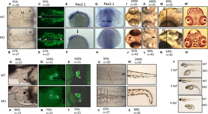
The morphological characteristics of zPnpo morphants. Zebrafish embryos of wild-type and transgenic lines injected with zPnpo MO at one- to two-cell stages were grown in embryo water and recorded for embryonic organogenesis and development. Tg(sox10:eGFP) embryos with/without MO injection were imaged at 18 hpf under dissecting light and fluorescent microscopes (AD) and also subjected to WISH analysis with riboprobes pax2.1(EH). Migratory neural crest cells (green fluorescent signals pointed by white arrows) were absent in the closing neural tube (dashed line) of wild-type larvae but remained in that of morphants. Lack or significantly reduced signal for pax2.1 (arrows) were also observed in zPnpo morphants. Both wild-type and zPnpo morphants at 2 dpf were imaged for brain region (IN). A crater-like opening was observed in diencephalic ventricle (arrows) and hindbrain (circled area) of zPnpo morphants. H&E-stained cryosections prepared from the head region [indicated by vertical lines in (N)] of 2-dpf morphants revealed a cavity (*), which was not observed in wild-type larvae (M’ and N’). Tg(cmlc2:eGFP) embryos with/without MO injection were imaged (OR) and video-recorded (S and T, the representative still frames from Suppl. 3–10) under a fluorescent microscope for heart development at 3 dpf. A tubing heart with incorrectly positioned ventricle and atrium were apparent in zPnpo morphants. The somite formation (U and V) and the development of trunk and tail (W and X) were recorded at 18 hpf and 3 dpf, respectively. The somites with chevron shape (solid line in U) can be seen in wild-type embryo. The full view of larvae at the indicated stages was imaged from the lateral view to show the overall morphology and size, which also revealed apparent body curvature for zPnpo morphants (Y). For the images to be taken, embryos/larvae of the indicated stages were removed from the embryo water and placed individually on a drop of methylcellulose for photographing. All images were taken with anterior to the left for lateral view except for brain (AD, EL) and somites (UV) (dorsal) and video recording (S and T) (ventral). WT, wild-type; MO, zPnpo morphants; F, forebrain ventricle; M, midbrain ventricle; H, hindbrain ventricle; nt, neural tube; V, ventricle; A, atrium; WISH, whole-mount in situ hybridization; H&E, hematoxylin and eosin.

Figure Data
Acknowledgments
This image is the copyrighted work of the attributed author or publisher, and ZFIN has permission only to display this image to its users. Additional permissions should be obtained from the applicable author or publisher of the image. Full text @ Front Pharmacol