
|
Fig. S2
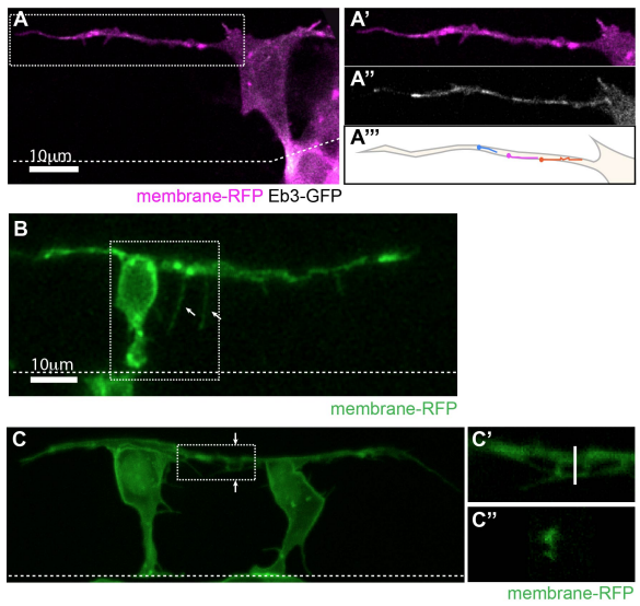
Basal protrusions contain dynamic microtubules, form filopodia and can overlap (related to Figure 1). A) T-shaped differentiating neuron with a long basal protrusion. White box marks the area of higher magnification shown in A’-A’’’). A’) Membrane-RFP. A’’) EB3-GFP (Movie 2). A’’’) Illustration of EB3-GFP comet trajectories during a 30 second timelapse period. B) T-shaped cell with filopodia (arrows) on basal protrusion (Movie 3). C) Two neurons differentiating 18µm apart with overlapping basal arms. C’) Higher magnification of white box in C). C’’) Cross-section of the basal protrusions at the line shown in C’). All images are projected images from confocal z-stacks. Dashed line shows the apical surface.
Reprinted from Developmental Cell, 49, Hadjivasiliou, Z., Moore, R.E., McIntosh, R., Galea, G.L., Clarke, J.D.W., Alexandre, P., Basal Protrusions Mediate Spatiotemporal Patterns of Spinal Neuron Differentiation, 907-919.e10, Copyright (2019) with permission from Elsevier. Full text @ Dev. Cell