
Image
|
Figure Caption
Fig. S1
–bubblebrain microglia are under osmotic stress –
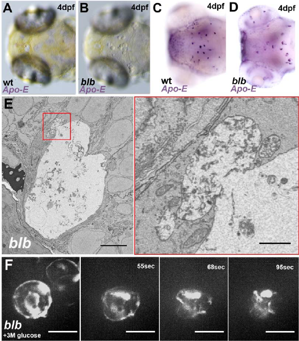
Related to Figure1 and Figure 2 (A-B) Bright field view of the dorsal side of a 4 dpf wild type (A) and blbNY007 (B) embryo. (C-D) WISH against Apo-E in a 4 dpf wild type (C) and blbNY007 (D) embryo. (E) Electron micrograph of a representative blbNY007 microglia at 3 dpf. Scale bar 5μm. Magnification of a fusion event. (F, movie 2) Injection of a 3M glucose solution in the brain of a blbNY007 embryo. Microglia labeled with Tg(spi1:Gal4,UAS:GFP). Time in seconds.
Acknowledgments
This image is the copyrighted work of the attributed author or publisher, and
ZFIN has permission only to display this image to its users.
Additional permissions should be obtained from the applicable author or publisher of the image.
Reprinted from Developmental Cell, 49(1), Villani, A., Benjaminsen, J., Moritz, C., Henke, K., Hartmann, J., Norlin, N., Richter, K., Schieber, N.L., Franke, T., Schwab, Y., Peri, F., Clearance by Microglia Depends on Packaging of Phagosomes into a Unique Cellular Compartment, 77-88.e7, Copyright (2019) with permission from Elsevier. Full text @ Dev. Cell