IMAGE

Figure 5

ID
ZDB-IMAGE-190723-600
Source
Figures for Gonzalez-Munoz et al., 2019
Image
Figure Caption

Figure 5

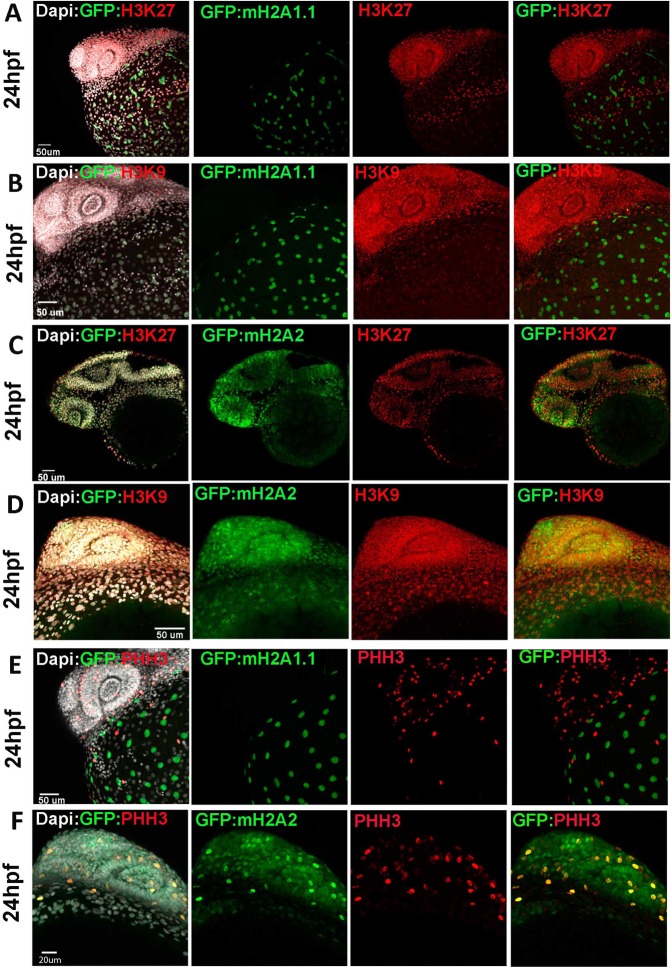
mH2A isoforms differential colocalization with heterochromatin and mitotic marks in zebrafish embryos at 24 hpf stage. Transgenic zf embryos expressing mH2A1:GFP-mH2A1.1 (A,B,E) or mH2A2:GFP-mH2A2 (C,D,F) at 24 hpf stage were analysed using immunohistochemistry to detect heterochromatin markers trimethyl Histone3 lysine K27 and K9 (H3K27me3 and H3K9me3) (A–D) or mitotic nuclei Phosphohistone H3 (pHH3) (E,F). DAPI was used for nucleus labelling. Confocal microscope image projection of 24 hpf transgenic fish. (A,B) mH2A1:GFP-mH2A1.1 embryo shows YSL localization of GFP positive cells and lack of colocalization with H3K27me3 (A,B) and H3K9m3 (C) throughout the embryo body. (C,D) mH2A2:GFP-mH2A2 embryo stained with heterochromatin markers H3K27me3 (D) and H3K9me3 (E) reveals areas of colocalization. (E,F) mH2A1:GFP-mH2A1.1 (E) or mH2A2:GFP-mH2A2 (F) with labelled pHH3 mitotic nuclei shows partial colocalization of GFP-mH2A2 isoform with mitotic nuclei. Each labelling assay was conducted in triplicate with n = 40–50 embryos/immunolabeling.

Acknowledgments
This image is the copyrighted work of the attributed author or publisher, and ZFIN has permission only to display this image to its users. Additional permissions should be obtained from the applicable author or publisher of the image. Full text @ Sci. Rep.