IMAGE

Figure 1

ID
ZDB-IMAGE-190723-2010
Source
Figures for Jo et al., 2013
Image
Figure Caption

Figure 1

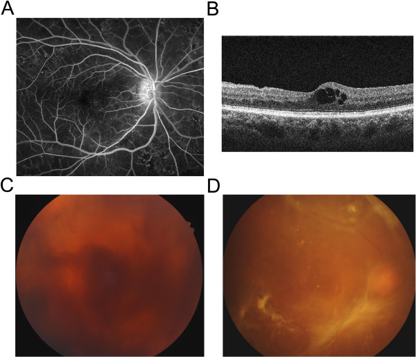
Pathologic changes in patients with DR. (A) Microaneurysm is one of the characteristic findings in patients with NPDR, demonstrated by tiny hyperfluorescent spots in the fluorescein angiography. (B) Disruption in neurovascular units and breakdown of inner BRB leads to ME in DR patients. Optical coherence tomography demonstrates cystoid ME. (C) In PDR patients, VH can occur from fragile new vessels. (D) Fibrovascular proliferation from retinal neovascularization forms diffuse tractional membrane, resulting in TRD.

Acknowledgments
This image is the copyrighted work of the attributed author or publisher, and ZFIN has permission only to display this image to its users. Additional permissions should be obtained from the applicable author or publisher of the image. Full text @ J. Biomed. Sci.