
Image
|
Figure Caption
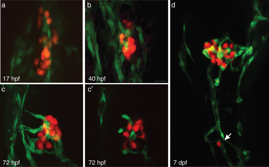
Fig. 1
The endocrine pancreas develops adjacent to vessels and is highly vascularized. (a–c) Confocal projections of the pancreatic islet at 17 hpf, 40 hpf, and 72 hpf in Tg(fli1:EGFP; insa:tagRFP); endothelial cells (green) and beta-cells (red). (c’) Confocal section of projection in (c). (d) Confocal projection of 7 dpf Tg(fli1:EGFP; insa:tagRFP) pancreas. Arrow indicates secondary islet.
Acknowledgments
This image is the copyrighted work of the attributed author or publisher, and
ZFIN has permission only to display this image to its users.
Additional permissions should be obtained from the applicable author or publisher of the image.
Full text @ Sci. Rep.