
|
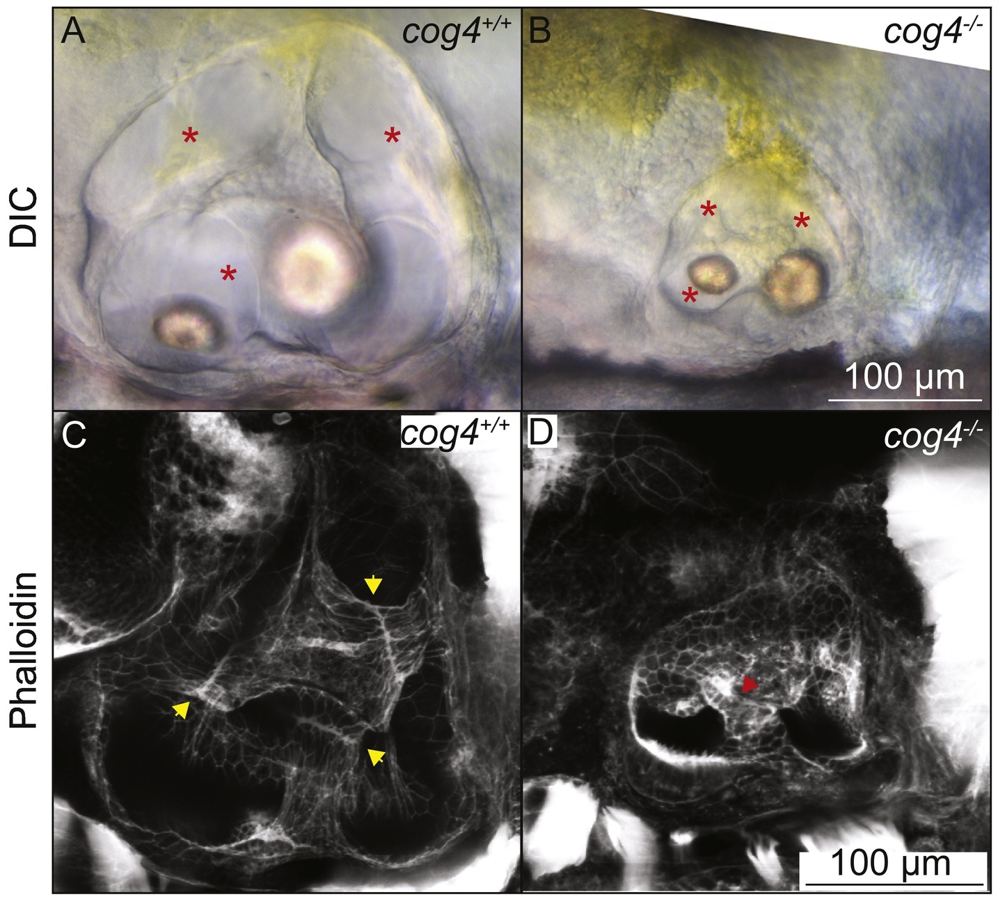
Fig. 1
The pillars do not form properly in cog4−/− mutants. Live images of the inner ear of cog4+/+ sibling (A, n = 34 larvae) and cog4−/− mutant larvae (B, n = 15 larvae). Red stars indicate the semicircular canals. Phalloidin staining of the inner ear of cog4+/+ sibling (C, n = 24 larvae) and cog4−/− mutant larvae (D, n = 25 larvae). Yellow arrowheads indicate the fusion plates that form in 100% (24 out of 24) of wild-type siblings (C). Red arrowhead points to the malformed pillar. One or more malformed pillars are observed in 84% (21 out of 25) of homozygous mutant larvae (D). Anterior to the left and dorsal to the top. 5 dpf.
Reprinted from Mechanisms of Development, 155, Clément, A., Blanco-Sánchez, B., Peirce, J.L., Westerfield, M., Cog4 is required for protrusion and extension of the epithelium in the developing semicircular canals, 1-7, Copyright (2018) with permission from Elsevier. Full text @ Mech. Dev.