
|
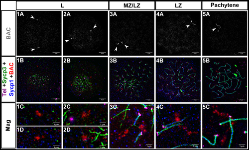
Fig. 3
1A-5A: Fluorescent in situ hybridization with a BAC probe (gray, white arrowheads) to a region ~10Mb from the telomere on chromosome 5. 1B-5B: Same images as 1A-5A plus telomeres (Tel; magenta), Sycp3 (green), and Sycp1 (blue); the BAC is in red. 1C-5C, 1D-2D: Magnifications (Mag) for each of the corresponding panels above. 1A-5B scale bar = 5 μm. Mag scale bar = 1 μm. Leptotene (L), mid to late zygotene (MZ/LZ); late zygotene (LZ). Note that images for panel series 3 and 5 are from a spreading procedure performed on a separate day from panel series 1, 2, and 4, and are included to illustrate a rare observation where BAC probes are located on axes where SC is extending (3C) and also where the BAC signal is highly elongated (5C).