
Image
|
Figure Caption
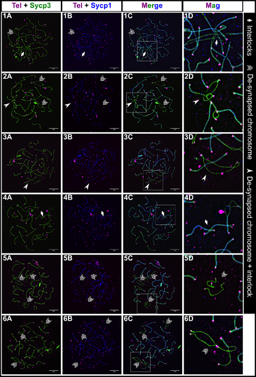
Fig. 5
Interlocks and de-synapsed chromosomes in male zebrafish meiosis I.
1A-6C: Examples of nuclear surface spreads showing interlocks (arrows) and de-synapsed chromosomes (triple arrows) at the late zygotene to pachytene stage where most chromosomes are synapsed. In some cases, de-synapsed chromosomes have another chromosome passing through them (arrowheads). 1D-6D: Magnification (Mag) images are from regions indicated by white boxes in the merge panels. Sycp3 (green), Sycp1 (blue), Telomeres (Tel; magenta). Scale bar = 5μm.
Figure Data
Acknowledgments
This image is the copyrighted work of the attributed author or publisher, and
ZFIN has permission only to display this image to its users.
Additional permissions should be obtained from the applicable author or publisher of the image.
Full text @ PLoS Genet.