
|
Fig. S2
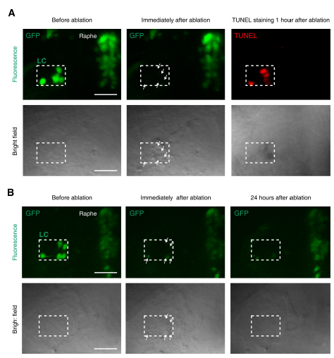
Confirmation of LC neuron death induced by two-photon laser-based ablation, Related to Figure 2.
(A) TUNEL signals in the ablated LC area in an ETvmat2:GFP larva. TUNEL staining was performed 1 hour after two-photon ablation. Top, fluorescent images showing GFP or TUNEL signal; bottom, bright field images showing bulb-like structures (white arrows). All images were obtained from the same larva. Scale: 50 μm.
(B) No GFP-positive cells in the ablated LC area in an ETvmat2:GFP larva 24 hours after two-photon ablation. Top, fluorescent images showing GFP signal; bottom, bright field images showing bulb-like structures (white arrows). All images were obtained from the same larva. Scale: 50 μm.