
Image
|
Figure Caption
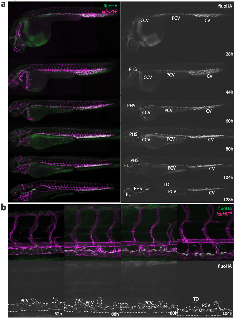
Fig. S8
FluoHA distribution through embryonic development. A. Whole-embryo view of fluorescent hyaluronic acid (fluoHA) distribution in kdrl:RFP transgenic embryos, 1h after injection. SECs, as identified through intracellular accumulation of fluoHA from 28hpf to at least 128hpf. From 104hpf, fluoHA uptake is also observed in lymphatic vessels, such as the thoracic duct (TD) and facial lymphatics (FL) B. Cellular view of fluoHA distribution in the trunk of kdrl:RFP transgenic embryos, 1h after injection. A gradual restriction of fluoHA accumulation to the PCV is observed between 52hpf and 104hpf.
Acknowledgments
This image is the copyrighted work of the attributed author or publisher, and
ZFIN has permission only to display this image to its users.
Additional permissions should be obtained from the applicable author or publisher of the image.
Full text @ ACS Nano