
|
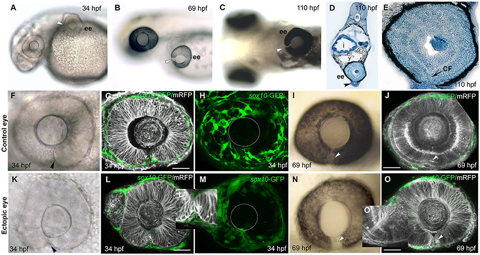
Fig. 3
Failure of choroid fissure fusion in eyes derived from optic vesicles with reduced POM. (A–C) Brightfield images of two host embryos at different stages after optic vesicle transplantation (at 12 ss, about 14 h pf), showing location of the ectopic eye on the yolk. Arrowheads indicate the open choroid fissure in the ectopic eyes. (D,E) Methylene blue stained sagittal sections at low (D) and high (E) resolution through an ectopic eye at 5 dpf. (F–O) Images of control (F–J) and ectopic (K–O) eyes imaged either in brightfield (F,K,I,N) or by confocal microscopy (G,H,L,M,J,O) showing cell membranes (mRFP, gray) and sox10:GFP labeled POM (green) at ages shown on the panels. Arrowheads indicate the choroid fissure. Asterisk in O' indicates retinal ganglion cell axons. The position of the lens is indicated with a white circle in (H) and (M). ee, ectopic eye; i, intestine; sc, spinal cord; y, yolk. Scale bars 50 μM.