
Fig. 3
|
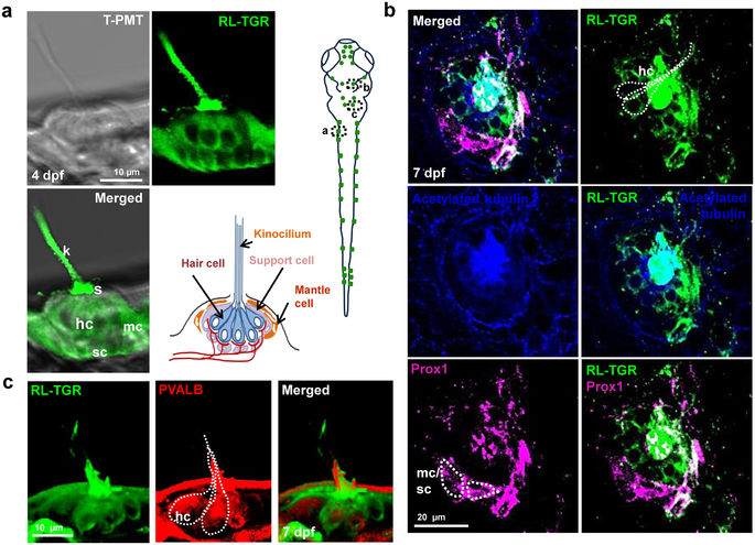
Fig. 3
RL-TGR is expressed in the neuromasts. (a) Confocal images of a single neuromast of the posterior lateral line system of wild type embryo at 4 dpf (lateral view). RL-TGR (green) staining signal was detected in hair cells (hc), support cells (sc), and mantle cells (mc) of the neuromast. The image on the bottom right represents the scheme of a neuromast illustrating different cells and their organization (modified after Ghysen and Dambly-Chaudiere, Current Opinion in Neurobiology, 2004)33. (b) Confocal images of a neuromast of wild type embryo (dorso-lateral view) at 7 dpf stained with RL-TGR (green, expression in the hair cells), acetylated tubulin (blue, expression in the cilia of the hair cells), and neuromast marker Prox1 (magenta, expression in the hair cells, supporting cells, and mantle cells). Colocalization of RL-TGR and acetylated tubulin suggests that RL-TGR is highly expressed in both kinocilia and cell bodies of the mechanosensory hair cells. RL-TGR colocalized with Prox1 in the stereocilia of the hair cells and supporting cells surrounding the neuromast. (c) 3D maximum intensity projection images of a neuromast of wild type embryo (lateral view) at 7 dpf stained with RL-TGR (green, expression in the hair cells), parvalbumin (PVALB) (red, expression in the hair cells). n > 50 embryos; schematic diagram of the 7 dpf larvae shows the location of different images; scale bars are shown in the images.