
|
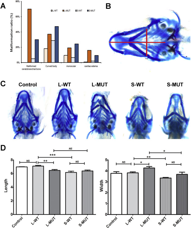
Fig. 6 In vivo analysis of two isoforms of zebrafish atp6v1h. A. The long and short zebrafish atp6v1h mRNA were injected into zebrafish embryos. After 7 days of injection, the ratios of malformed ceratobranchial bone, curved body, monocular and cardiac edema were compared among long mRNA (L-WT, L-MUT) and short mRNA (S-WT, S-MUT). The mutant group showed a higher malformation percent that the wild type in one representative experiment. And the short isoform showed an obvious lower ratio of malformed ceratobranchial bone than the longer one. B ~ D. Comparison of zebrafish heads. The embryos were collected and stained with alcian blue/alizarin red after 7 days of injection. Both long and short variants with c.G1092T_A1093T (NM_001291708) caused malformation changes in craniofacial bone. The short atp6v1h mRNA reduced the length and width of the zebrafish. B. Red lines represent the length (from the front of Meckel's cartilage to the connections of two 5th ceratobranchial structures) and width (distance between the outermost vertex of the left and right ceratohyal) for the calculations. C. Represent images in each group. D. Comparison tables. n ≥ 20.WT: wild type, L: long variant, S: short variant. MUT: Double mutants G1092T_A1093T. NS: not significant. *: P < 0.05; **: P < 0.01; ***:P < 0.001. (For interpretation of the references to colour in this figure legend, the reader is referred to the web version of this article.)
Reprinted from Gene, 638, Zhao, W., Zhang, Y., Yang, S., Hao, Y., Wang, Z., Duan, X., Analysis of two transcript isoforms of vacuolar ATPase subunit H in mouse and zebrafish, 66-75, Copyright (2018) with permission from Elsevier. Full text @ Gene