
Image
|
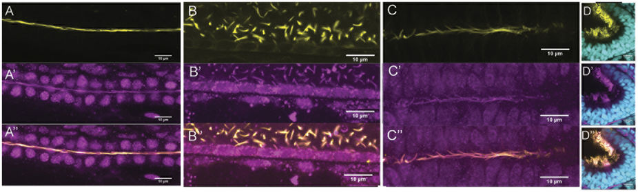
Figure Caption
Fig. 2
Rsph9 protein is enriched in cilia.
Wildtype embryos were stained with an anti-Rsph9 antibody (magenta) and acetylated α-tubulin (yellow). (A) Pronephric duct. (B) Ventral spinal cord. (C) Ventral midbrain. (D) Olfactory pit, nuclei are visualized with DAPI (cyan). All images are shown with anterior to the left. (A,B) Are lateral mounts (dorsal at the top).
Figure Data
Acknowledgments
This image is the copyrighted work of the attributed author or publisher, and
ZFIN has permission only to display this image to its users.
Additional permissions should be obtained from the applicable author or publisher of the image.
Full text @ Sci. Rep.