
|
Fig. 1
HSPC Localization in Zebrafish CHT, and RNA-Seq Analysis
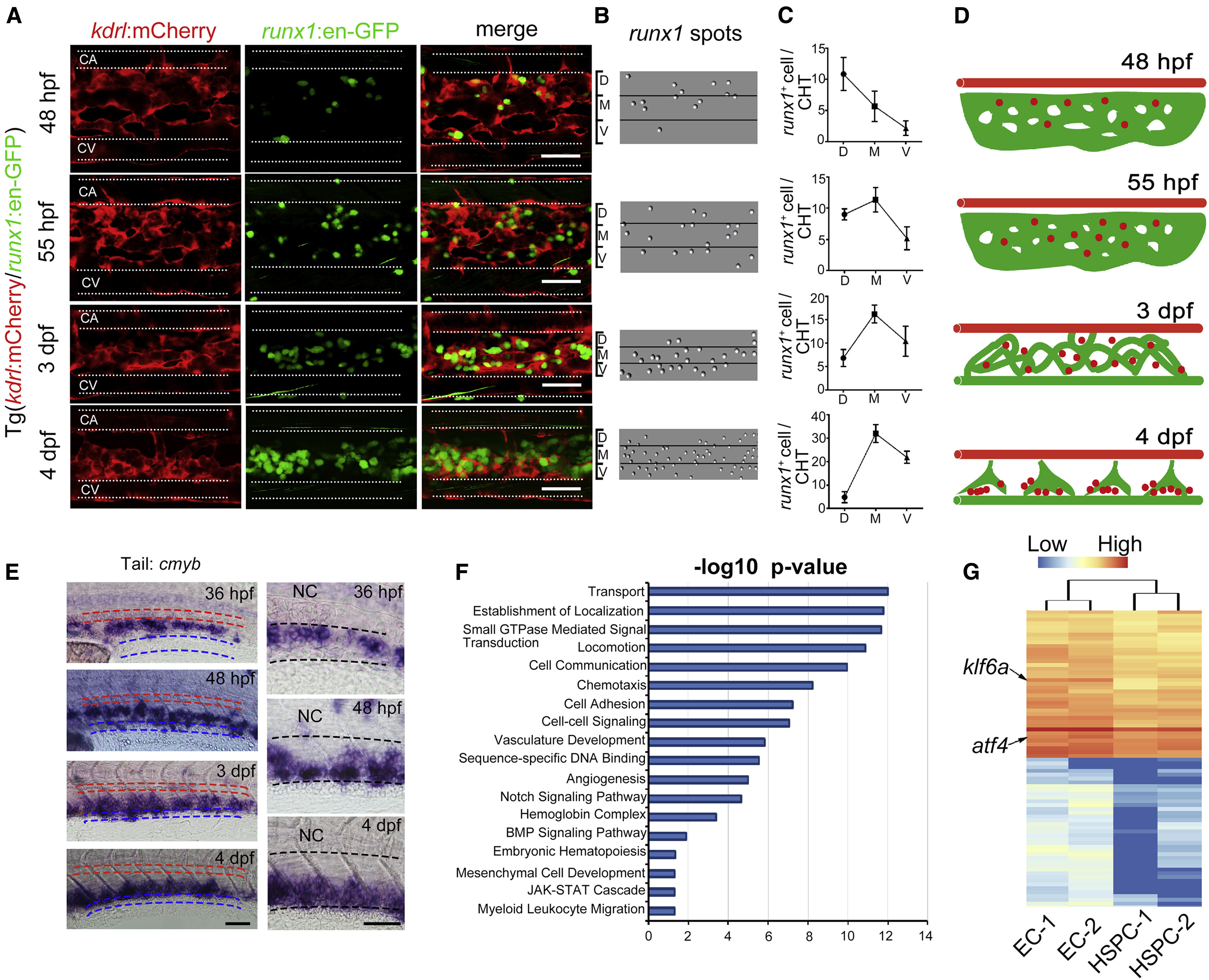
(A) In vivo imaging of Tg (kdrl:mCherry/runx1:en-GFP) showing the detailed localization of HSPCs in the CHT during dynamic developmental processes. At 48 hpf, runx1+ cells mainly localized next to the ventral wall of caudal artery and then migrated into the venous plexus at 55 hpf. At a later stage, i.e., 3 dpf and 4 dpf, runx1+ cells seeded the dorsal wall of caudal vein in the CHT. The white dashed lines indicate the outlines of caudal artery (CA) and caudal vein (CV), respectively. Scale bars, 30 μm.
(B) The runx1 spots in the CHT were achieved from runx1:en-GFP+ cells in (A) via imaging by Imaris processing.
(C) The statistical data of runx1 spots in D/M/V region during developmental stages.
(D) The model of dynamic development of CVP and associated HSPCs. The red color marks caudal artery, the green color marks CVP, and red dots mark HSPCs.
(E) Expression of cmyb in the CHT at successive developmental stages. The red dashed lines mark the caudal artery and blue dashed lines mark the main caudal vein. The right panels are magnified images of cmyb expression in the CHT. The black dashed lines indicate the boundary between the caudal artery and vein. NC, notochord. Scale bars, 30 μm.
(F) DAVID gene ontology (GO) analysis of genes significantly enriched in ECs versus HSPCs.
(G) Heatmap displays selected highly expressed TFs in ECs of 52 hpf from the GO analysis. The arrows indicate two genes, atf4 and klf6a, respectively.
See also Figure S1.
Reprinted from Developmental Cell, 42(4), Xue, Y., Lv, J., Zhang, C., Wang, L., Ma, D., Liu, F., The Vascular Niche Regulates Hematopoietic Stem and Progenitor Cell Lodgment and Expansion via klf6a-ccl25b, 349-362.e4, Copyright (2017) with permission from Elsevier. Full text @ Dev. Cell