
|
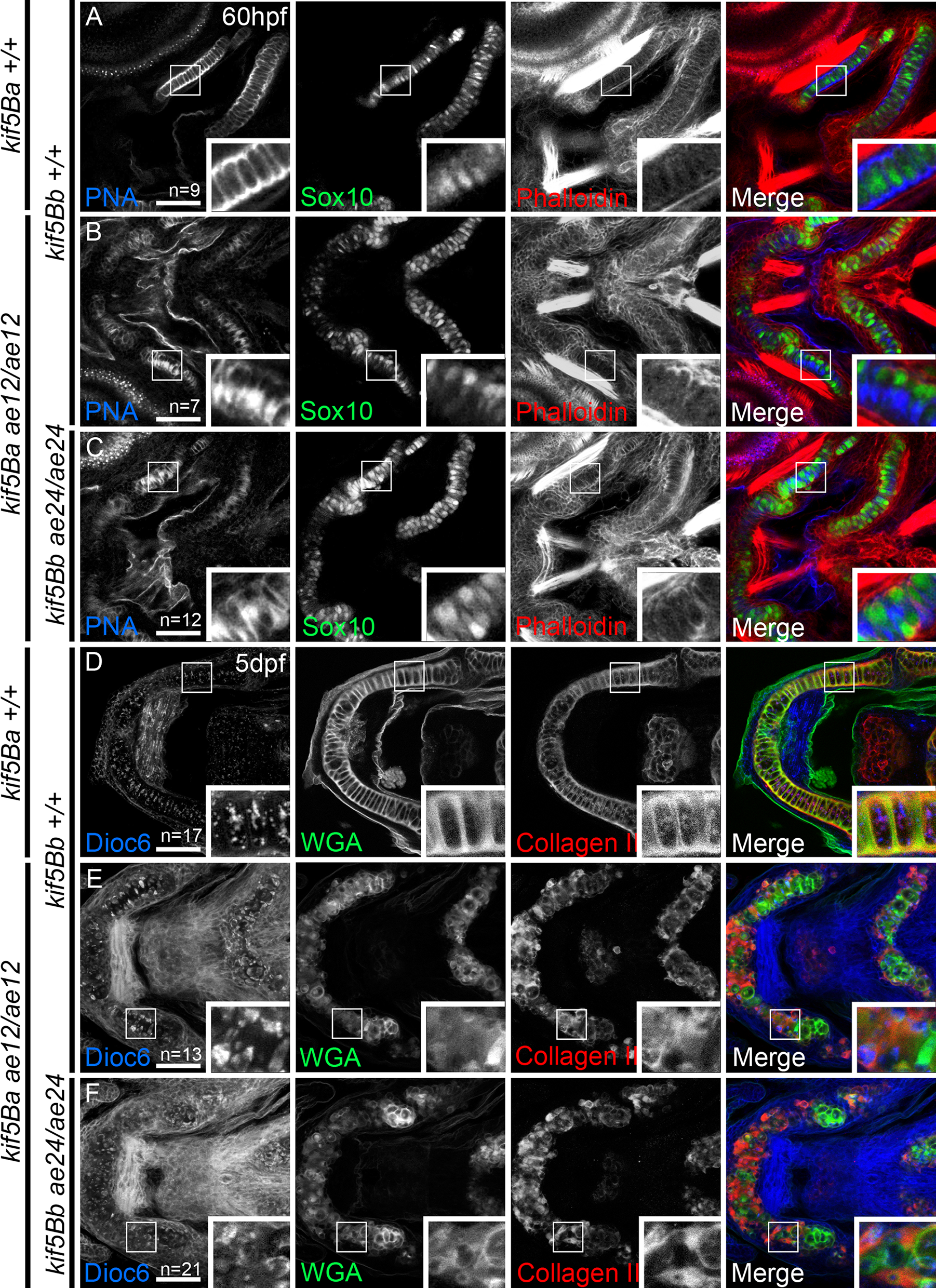
Fig. 2
Loss of kif5B impairs chondrocyte secretion.
A-C) confocal images of Wt (A), single kif5Baae12/ae12 mutants (B) and kif5Baae12/ae12Kif5Bbae24/ae24 (C) at 60 hpf. Chondrocytes (Sox10:GFP positive cells) secrete proteins positive for peanut agglutinin (PNA, blue) homogeneously. Secretion in single (B) and compound (C) mutants is deficient. Stacking defects are evident in kif5Baae12/ae12Kif5Bbae24/ae24 (C). D-F) Confocal images of Wt (D), single kif5Baae12/ae12 mutants (E) and kif5Baae12/ae12Kif5Bbae24/ae24 (F) at 5 dpf. In Wt (D) Di0C6 (blue) forms variably-sized cellular aggregates. Wheat Germ agglutinin (WGA, green) labels secreted proteins and II-II6B3 marks Collagen II (red) an abundant protein in cartilage. Both are homogeneously secreted in Wt and overlap within ECM (insets in D), although some overlap occurs between Collagen II and Di0C6 (insets in D). In single (E) and compound (F) mutants, Di0C6 forms large patches. Secretion is polarized with little colocalization within ECM (insets in E and F), large patches of Di0C6 colocalize with Collagen II, and cell stacking is perturbed (E, F). Scale bar: 50μm.