
|
Fig. S3
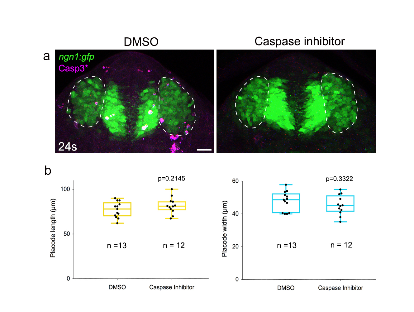
Proliferation and associated cell size changes are not involved in OP morphogenesis. (a) Spatial distribution of PH3+ dividing cells in ngn1:gfp placodes at 16s and 24s. Comparison with a uniform distribution using a Chi2 test gave a p value>0.05, showing the uniform distribution of dividing cells. (b) Graphs showing the decrease in PH3+ cells at 16s, and the decrease in GFP+ cell numbers in OPs at 24s observed in embryos treated with the anti-proliferation drugs hydroxyurea and aphidicolin (HUA), or DMSO. (c) ngn1 :gfp embryos incubated with HUA or DMSO from 12s to 24s, showing no overt OP morphogenesis defects upon HUA treatment. Dotted lines surround the two paired OPs. (d) Quantification of OP length and width at 24s in embryos treated with HUA or DMSO controls. In (b) and (c) n indicates the number of analysed placodes in each condition. p values: unpaired two-tailed t-tests. (e,f) ngn1:gfp embryos injected with mbCherry mRNA were fixed and stained with DAPI at 12s and 24s. Individual cell bodies were reconstructed in 3D using the mbCherry staining. Right panels show examples of reconstructed cells, corresponding to the cells highlighted in magenta in the left panels. (g) The 3D cell reconstruction allowed to quantify cell volume, cell height (along the DV axis) and cell area (maximum XY area) at 12s (n= 15 cells from N=6 placodes) and 24s (n= 15 from N=4 placodes). p values: unpaired two-tailed t-tests. Cell volume is smaller at 24s than at 12s, which is mostly due to a smaller cell size in the XY plan, rather than a reduction of cell height in the DV (Z) axis. (h) Nucleus area correlates with cell area, and can thus be used as a readout for cell size. (i) Instance of a placodal cell undergoing division, showing the decreased area in the two daughter cells, compared with the mother cell. U) Dynamic measurement of areas of placode cell nuclei in live imaging experiments, showing that cell division coincides with a reduction in the area of nuclei (averaged on n=13 cells). (k) DAPI staining on 24s ngn1:gfp embryos incubated with HUA (rigth) or DMSO (left), showing placodes with similar size but bigger cells in HUA-treated embryos. (I) Quantification of OP volume and average cell volumes in embryos treated with HUA or DMSO. n indicates the number of analysed placodes in each condition p values: unpaired two-tailed t-tests. Scale bars: 25 µm except in I: 5 µm.