
|
Fig. 14
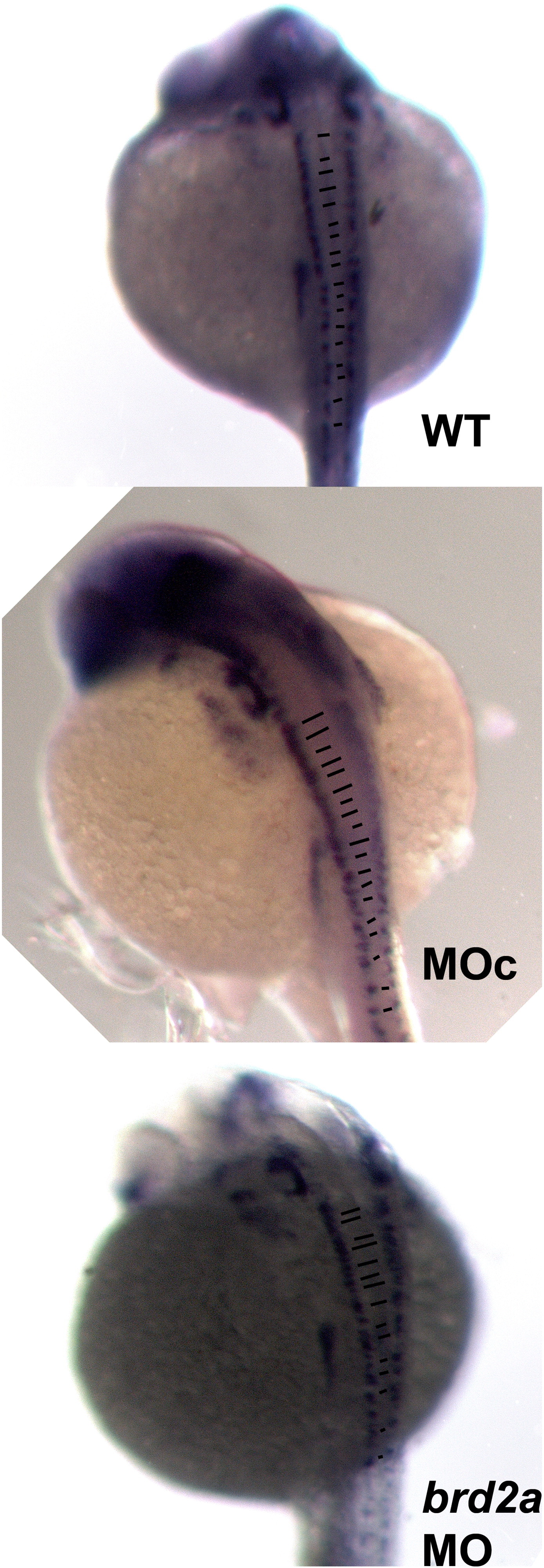
brd2aMO morphants exhibit disrupted patterning of hindbrain and spinal interneurons. Embryos from pax2a in situ to RNA experiments were examined to visualize various pax2a (+) cells along the A-P axis. Labeling of hindbrain and spinal interneurons by pax2a reveals A-P patterning of paired neurons on either side of the neural tube in wildtype uninjected (WT), control MOc-injected (MOc) and brd2aMO1-injected (brd2aMO) embryos. In wildtype embryos, neurons are perfectly matched 97.1% of the time, with only 2.9% showing 2–3 mismatched pairs (A); similar results are observed in the morpholino control MOc group (B). In contrast, neurons in brd2a knockdown embryos (C) are normally matched only 17.2% of the time, with 48.3% showing 2–3 mismatches, and 34.5% showing an unrecognizable pattern (p < 0.0001, chi square contingency).
Reprinted from Mechanisms of Development, 146, Murphy, T., Melville, H., Fradkin, E., Bistany, G., Branigan, G., Olsen, K., Comstock, C.R., Hanby, H., Garbade, E., DiBenedetto, A.J., Knockdown of epigenetic transcriptional co-regulator Brd2a disrupts apoptosis and proper formation of hindbrain and midbrain-hindbrain boundary (MHB) region in zebrafish, 10-30, Copyright (2017) with permission from Elsevier. Full text @ Mech. Dev.