
Image
|
Figure Caption
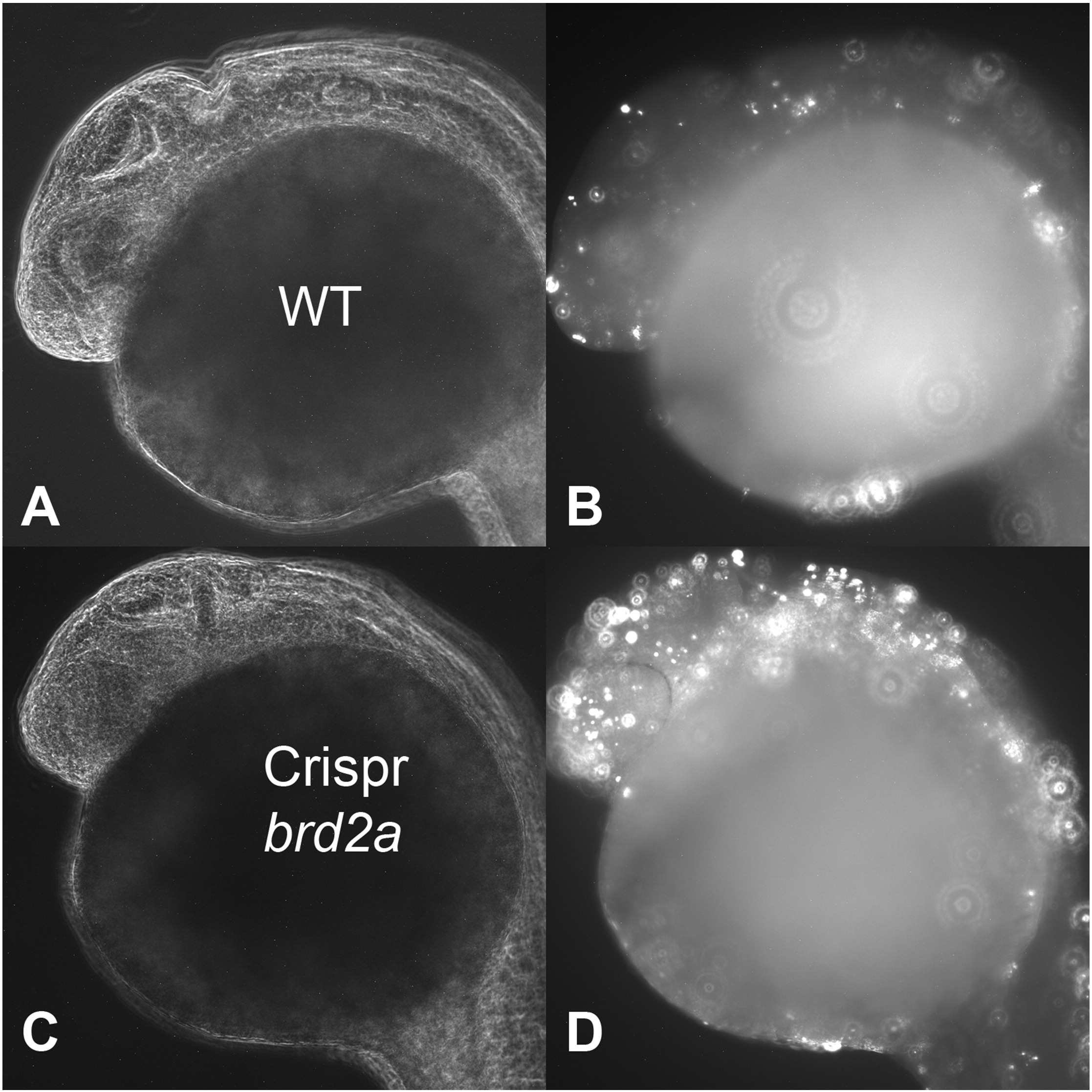
Fig. 7
brd2a Crispr/Cas9 injections corroborate brd2aMO morphological and cell death defects.
One cell stage embryos were co-injected with brd2a crRNA/tracrRNA and Cas9 mRNA, assayed by TUNEL for apoptotic nuclei at prim 5 stage, and examined by laser scanning confocal microscopy. At least 10–15 embryos from two clutches were examined. Light and darkfield images of wildtype uninjected (A,B) and brd2a Crispr/Cas9 injected (C,D) representative embryos are shown. Levels of cell death are significantly increased in brd2a Crispr/Cas9 treated samples. Refer to D for quantitative analysis.
Figure Data
Acknowledgments
This image is the copyrighted work of the attributed author or publisher, and
ZFIN has permission only to display this image to its users.
Additional permissions should be obtained from the applicable author or publisher of the image.
Reprinted from Mechanisms of Development, 146, Murphy, T., Melville, H., Fradkin, E., Bistany, G., Branigan, G., Olsen, K., Comstock, C.R., Hanby, H., Garbade, E., DiBenedetto, A.J., Knockdown of epigenetic transcriptional co-regulator Brd2a disrupts apoptosis and proper formation of hindbrain and midbrain-hindbrain boundary (MHB) region in zebrafish, 10-30, Copyright (2017) with permission from Elsevier. Full text @ Mech. Dev.