
Image
|
Figure Caption
Fig. S1
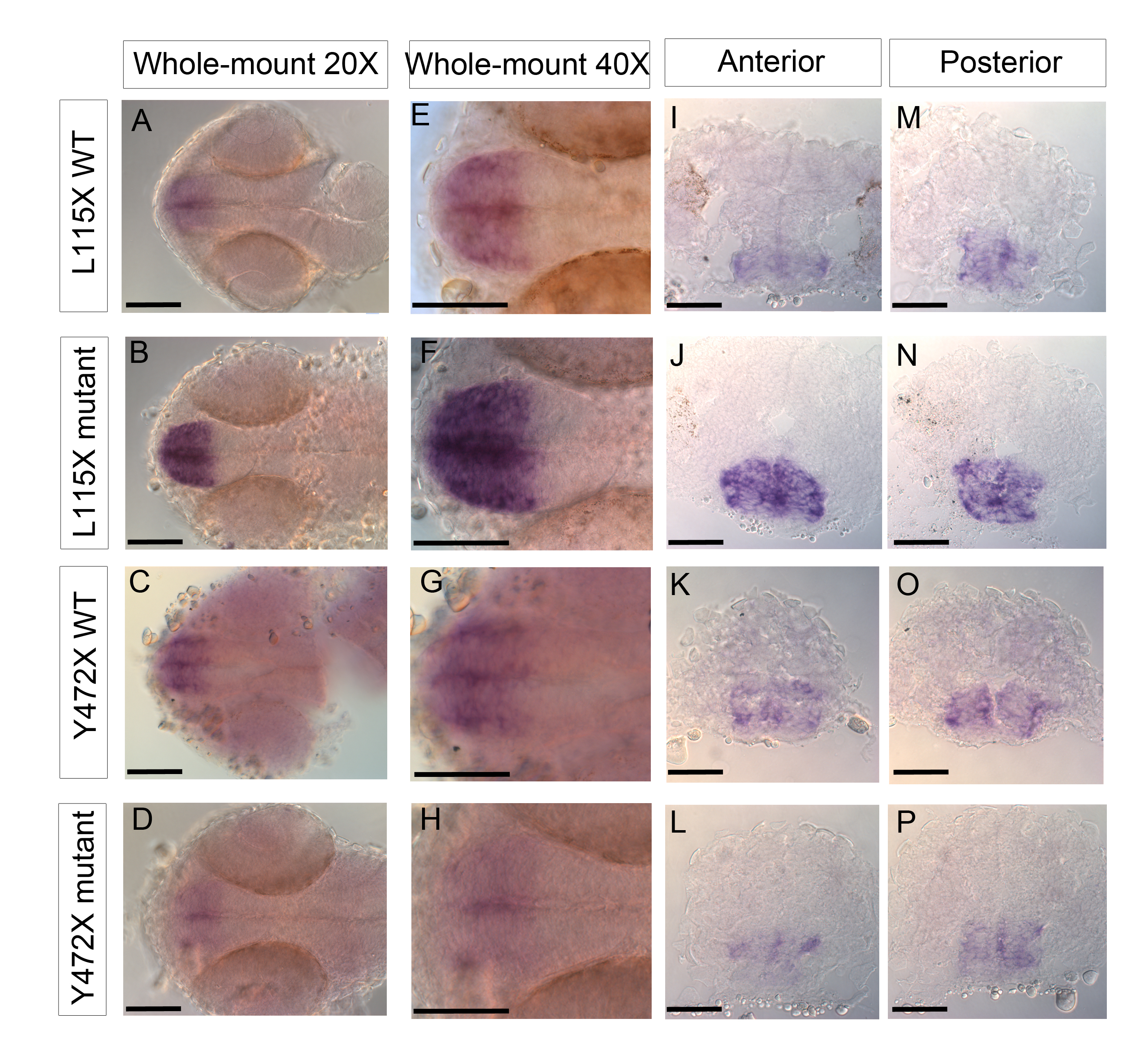
Expression of rx3 by in situ hybridization in 24 hpf disc1 embryos. (A-H) Ventral whole-mount view of rx3 in the developing hypothalamus, showing increased expression in L115X mutants (B,F) compared to wild types (A,E) and reduced expression in Y472X mutants (D,H) compared with wild type embryos (C,G). (A-D) are 20X magnification, (E-H) are 40X magnification. Anterior left. (I-P) Representative transverse sections through the anterior (I-L) and posterior (M-P) regions of the developing hypothalamus in L115X (I, J, M, N) and Y472X (K, L, O, P) embryos. Scale bars: 50 μm.
Acknowledgments
This image is the copyrighted work of the attributed author or publisher, and
ZFIN has permission only to display this image to its users.
Additional permissions should be obtained from the applicable author or publisher of the image.
Full text @ Hum. Mol. Genet.