
|
Fig. S3
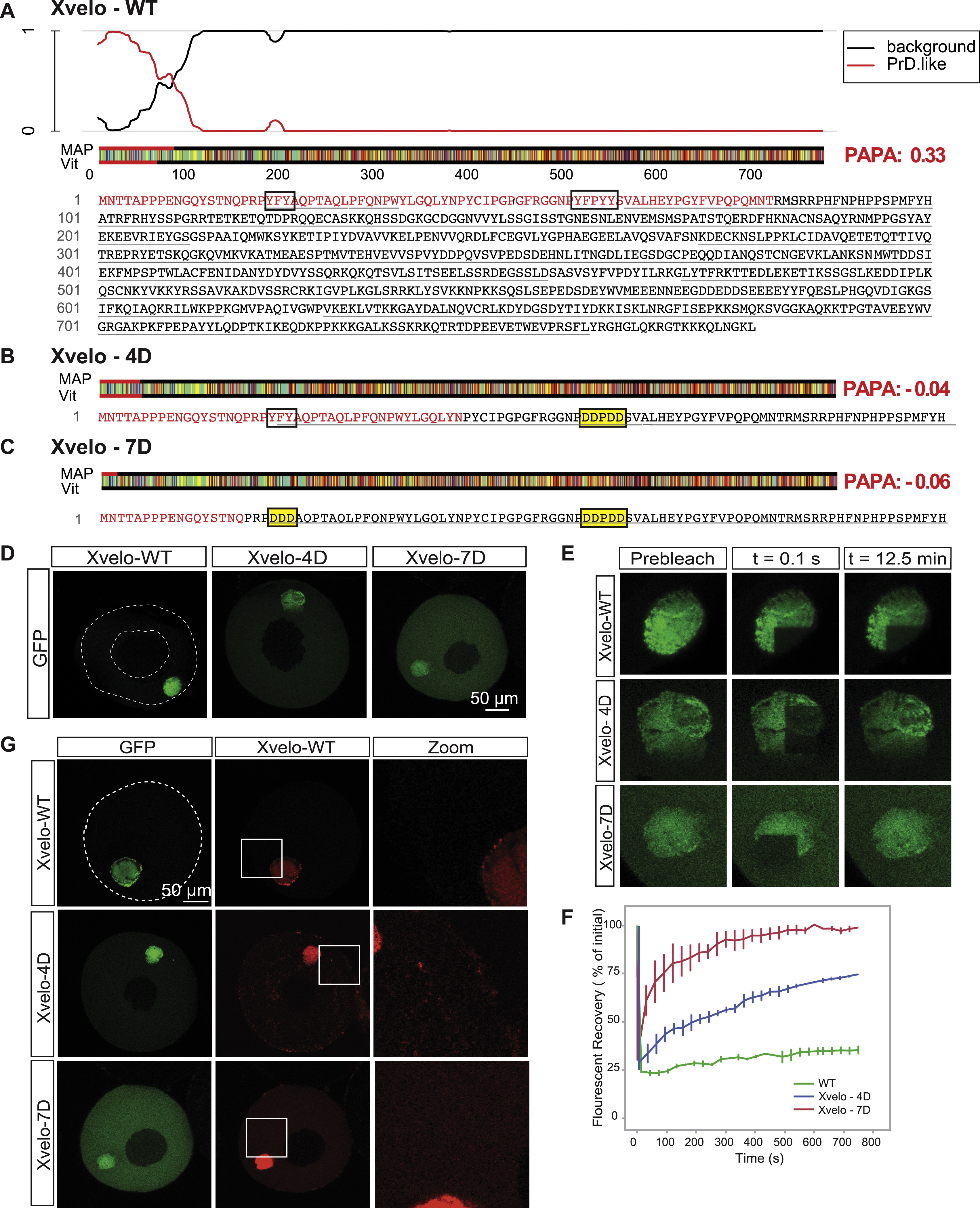
Xvelo Has a Prion-like Domain Predicted by PLAAC and PAPA, and Full-Length Mutants of Xvelo Recapitulated F1 Fragment Kinetics, Related to Figure 3
(A) Xvelo has an N-terminal prion-like domain predicted by PLAAC, and PAPA prion prediction programs. PLAAC results are shown here, and PAPA scores are highlighted in red at the right handside (Toombs et al., 2012 ; Lancaster et al., 2014). The aminoacid sequence of the protein is color coded according to Lancaster et al., 2014. The aromatic residues which are mutated are boxed in for ease of visualization. PrD.like: prion-like domain. Vit and Map correspond to the maximum length of consecutive PrD state in Viterbi or MAP parses.
(B) Mutating the aromatic residues in the prion-like domain of Xvelo to charged residues, YFPYY into DDPDD, stopped Xvelo score positive in PAPA prion detection algorithm. 4D mutant scored –0.04 in PAPA, and the aggregation-prone region detected by PLAAC decreased more than half in length.
(C) Additional mutations of aromatic residues in (B) to charged residues, YFY to DDD, further eliminated the detection of Xvelo by prion-prediction algorithms. The PAPA score decreased to –0.059, and PLAAC detected few aminoacids at the N-terminal as aggregation-prone.
(D) Full-length Xvelo-GFP, Xvelo-4D-GFP and Xvelo-7D-GFP mRNAs were microinjected into oocytes, incubated overnight in OCM, and imaged by scanning confocal microscopy. Note the 4D mutation has a stronger effect on the localization of the F1 fragment than the full-length protein (Figure 3D).
(E) A region corresponding to a quarter of a Balbiani body was photobleached in the oocytes shown in (D), and the fluorescence recovery of GFP was monitored in every 30 s for 12.5 min.
(F) Quantification of the oocytes photobleached in (E) and two other biological replicates. SEs were plotted.
(G) mRNAs encoding for full-length Xvelo-mCherry, Xvelo-GFP, Xvelo-4D-GFP and Xvelo-7D-GFP were co-injected into oocytes, and imaged after overnight incubation.
Reprinted from Cell, 166, Boke, E., Ruer, M., Wühr, M., Coughlin, M., Lemaitre, R., Gygi, S.P., Alberti, S., Drechsel, D., Hyman, A.A., Mitchison, T.J., Amyloid-like Self-Assembly of a Cellular Compartment, 637-50, Copyright (2016) with permission from Elsevier. Full text @ Cell